Kaverina I, Krylyshkina O, Small J V
Institute of Molecular Biology, Austrian Academy of Sciences, A-5020 Salzburg, Austria.
J Cell Biol. 1999 Sep 6;146(5):1033-44. doi: 10.1083/jcb.146.5.1033.
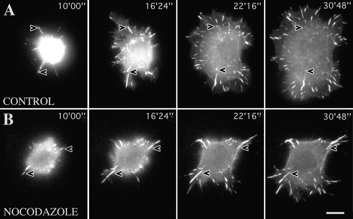
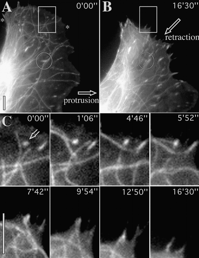
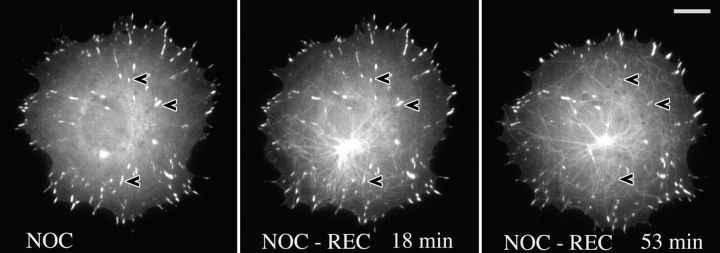
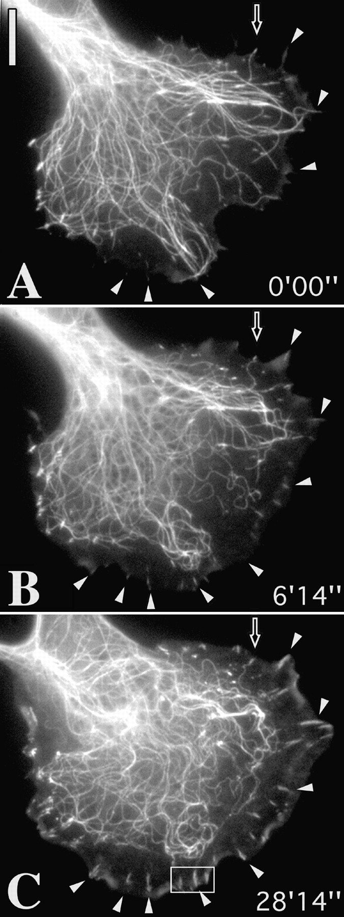
We recently showed that substrate contact sites in living fibroblasts are specifically targeted by microtubules (Kaverina, I., K. Rottner, and J.V. Small. 1998. J. Cell Biol. 142:181-190). Evidence is now provided that microtubule contact targeting plays a role in the modulation of substrate contact dynamics. The results are derived from spreading and polarized goldfish fibroblasts in which microtubules and contact sites were simultaneously visualized using proteins conjugated with Cy-3, rhodamine, or green fluorescent protein. For cells allowed to spread in the presence of nocodazole the turnover of contacts was retarded, as compared with controls and adhesions that were retained under the cell body were dissociated after microtubule reassembly. In polarized cells, small focal complexes were found at the protruding cell front and larger adhesions, corresponding to focal adhesions, at the retracting flanks and rear. At retracting edges, multiple microtubule contact targeting preceded contact release and cell edge retraction. The same effect could be observed in spread cells, in which microtubules were allowed to reassemble after local disassembly by the application of nocodazole to one cell edge. At the protruding front of polarized cells, focal complexes were also targeted and as a result remained either unchanged in size or, more rarely, were disassembled. Conversely, when contact targeting at the cell front was prevented by freezing microtubule growth with 20 nM taxol and protrusion stimulated by the injection of constitutively active Rac, peripheral focal complexes became abnormally enlarged. We further found that the local application of inhibitors of myosin contractility to cell edges bearing focal adhesions induced the same contact dissociation and edge retraction as observed after microtubule targeting. Our data are consistent with a mechanism whereby microtubules deliver localized doses of relaxing signals to contact sites to retard or reverse their development. We propose that it is via this route that microtubules exert their well-established control on cell polarity.
我们最近发现,活成纤维细胞中的底物接触位点是微管的特异性靶向目标(卡韦里纳,I.,K. 罗特纳,和 J.V. 斯莫尔。1998 年。《细胞生物学杂志》142:181 - 190)。现在有证据表明,微管接触靶向在底物接触动力学的调节中起作用。这些结果来自于铺展和极化的金鱼成纤维细胞,其中微管和接触位点通过与 Cy - 3、罗丹明或绿色荧光蛋白偶联的蛋白质同时可视化。与对照组相比,在诺考达唑存在下铺展的细胞,接触的周转受到阻碍,并且在细胞体下方保留的黏附在微管重新组装后解离。在极化细胞中,在突出的细胞前端发现小的粘着斑复合物,在收缩侧翼和后端发现较大的黏附,对应于粘着斑。在收缩边缘,多个微管接触靶向先于接触释放和细胞边缘收缩。在铺展细胞中也能观察到相同的效果,在这些细胞中,通过向一个细胞边缘施加诺考达唑使其局部解聚后,微管重新组装。在极化细胞突出的前端,粘着斑复合物也成为靶向目标,结果其大小要么保持不变,要么更少见地被拆解。相反,当用 20 nM 紫杉醇冻结微管生长来阻止细胞前端的接触靶向,并通过注射组成型激活的 Rac 刺激突起时,周边粘着斑复合物异常增大。我们进一步发现,将肌球蛋白收缩性抑制剂局部应用于带有粘着斑的细胞边缘,会诱导与微管靶向后观察到的相同的接触解离和边缘收缩。我们的数据与一种机制一致即微管将局部剂量的松弛信号传递到接触位点以延缓或逆转其发展。我们提出,微管正是通过这条途径对细胞极性发挥其已确立的控制作用。