Taraszka K S, Higgins J M, Tan K, Mandelbrot D A, Wang J H, Brenner M B
Lymphocyte Biology Section, Division of Rheumatology, Immunology and Allergy, Department of Internal Medicine, Bringham and Women's Hospital, Boston, MA 02115, USA.
J Exp Med. 2000 May 1;191(9):1555-67. doi: 10.1084/jem.191.9.1555.
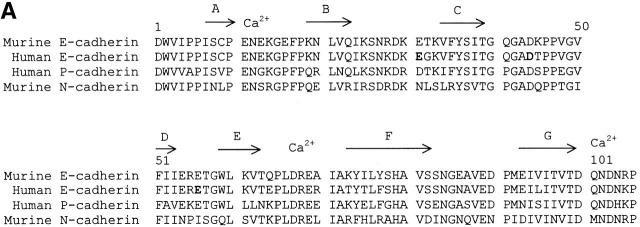
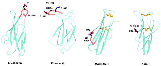
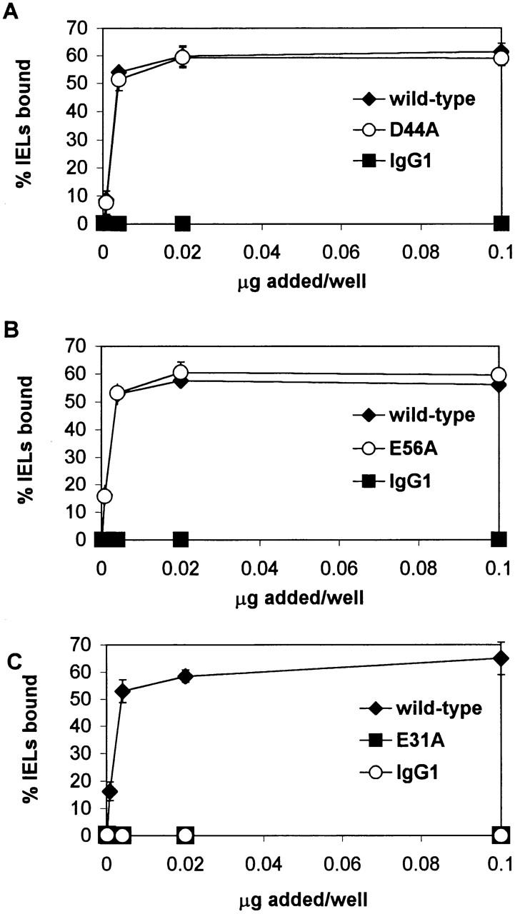
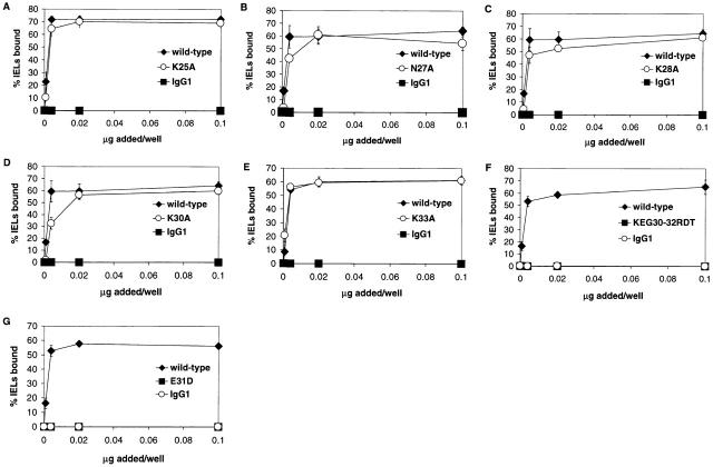
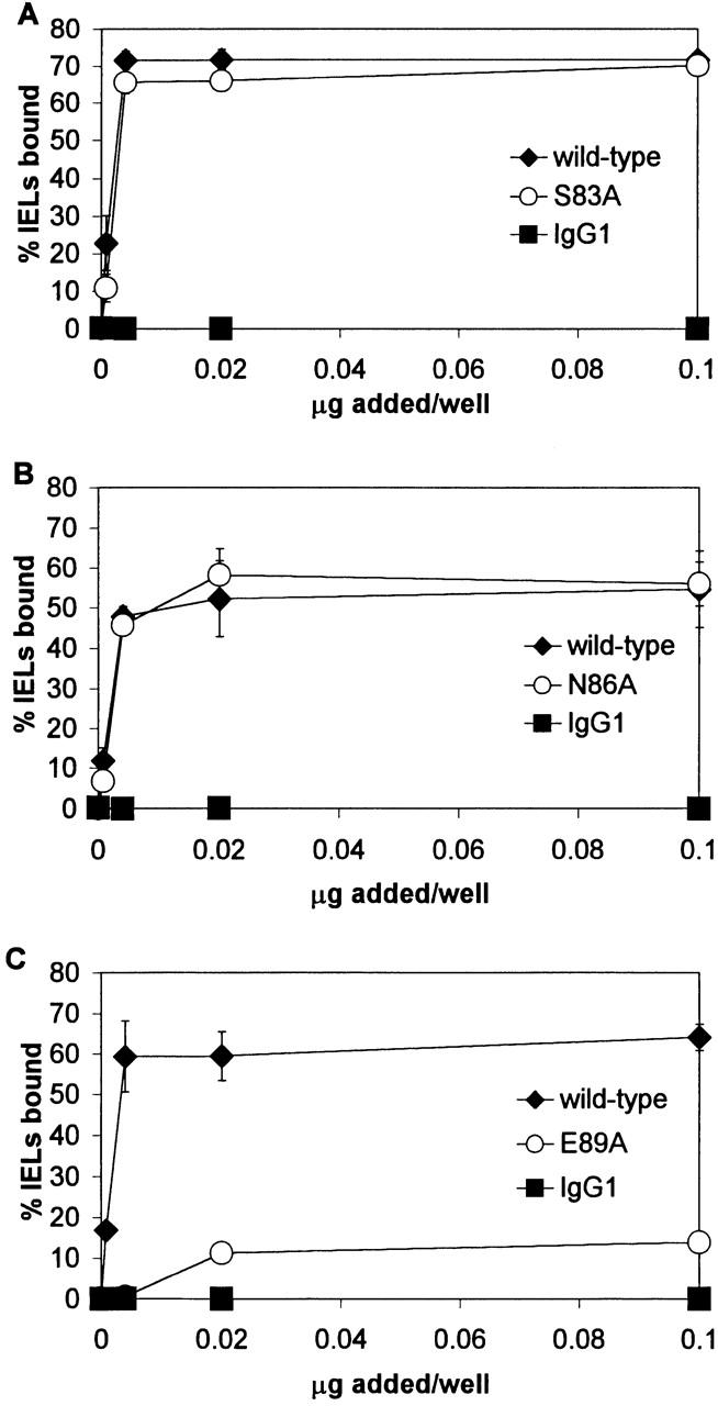
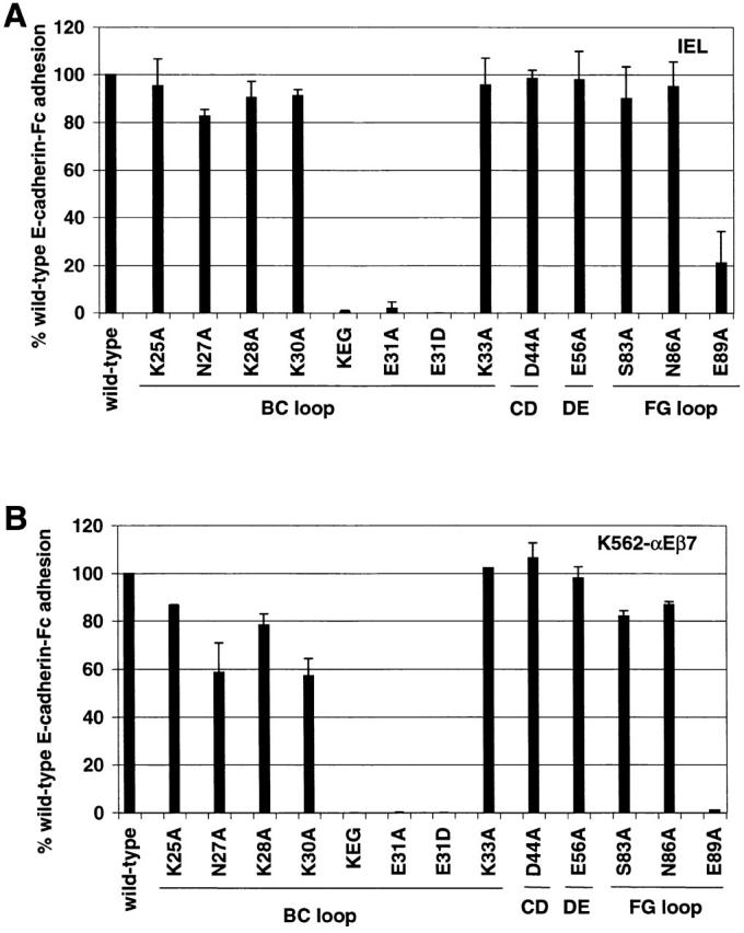
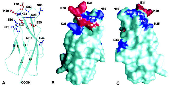
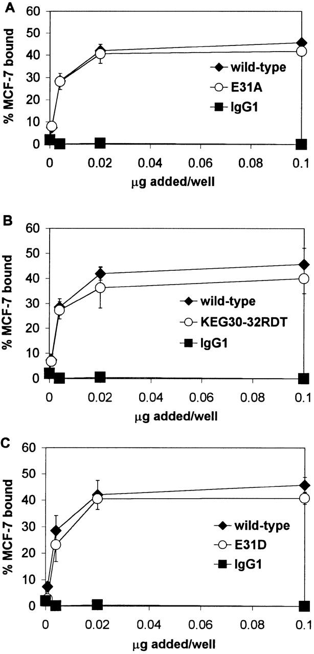
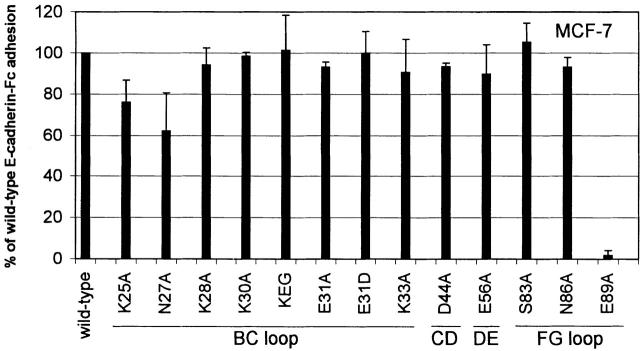
Cadherins are expressed in tissue-restricted patterns and typically mediate homophilic adhesion. Cadherins also mediate lymphocyte adhesion, providing the opportunity for lymphocyte attachment to parenchymal cells. The best characterized example of lymphocyte adhesion to a tissue-specific cell adhesion molecule, as opposed to a vascular endothelial adhesion molecule, is the interaction between integrin alpha(E)beta(7) on intraepithelial lymphocytes and E-cadherin on epithelial cells. However, the molecular basis for an integrin-cadherin interaction is not well defined. Realization that the cadherin domain adopts a topology similar to the immunoglobulin (Ig) fold suggested that integrin recognition of E-cadherin might be similar to recognition of Ig superfamily ligands. Thus, we modeled domain 1 of human E-cadherin and studied the role of solvent-exposed loops that connect Ig-like core-forming beta strands. Mutational analyses localized the integrin alpha(E)beta(7) recognition site to the top of domain 1 at the face formed by the BC and FG loops, a site distinct from the region recognized in intercellular adhesion molecule (ICAM)-1, -2, and -3, mucosal addressin cell adhesion molecule 1 (MAdCAM-1), vascular cell adhesion molecule 1 (VCAM-1), and fibronectin by their integrin ligands. Moreover, the integrin alpha(E)beta(7) binding site is distinct from the homophilic binding site on E-cadherin. These studies provide a conceptual basis for integrin-cadherin binding and extend the model that an Ig-like fold can serve as a scaffold for recognition.
钙黏蛋白以组织限制性模式表达,通常介导同嗜性黏附。钙黏蛋白还介导淋巴细胞黏附,为淋巴细胞与实质细胞的附着提供了机会。与血管内皮黏附分子不同,淋巴细胞与组织特异性细胞黏附分子黏附的最典型例子是上皮内淋巴细胞上的整合素α(E)β(7)与上皮细胞上的E-钙黏蛋白之间的相互作用。然而,整合素-钙黏蛋白相互作用的分子基础尚未明确界定。认识到钙黏蛋白结构域采用类似于免疫球蛋白(Ig)折叠的拓扑结构,提示整合素对E-钙黏蛋白的识别可能类似于对Ig超家族配体的识别。因此,我们对人E-钙黏蛋白的结构域1进行了建模,并研究了连接Ig样核心形成β链的溶剂暴露环的作用。突变分析将整合素α(E)β(7)识别位点定位到结构域1顶部由BC环和FG环形成的表面,该位点不同于细胞间黏附分子(ICAM)-1、-2和-3、黏膜地址素细胞黏附分子1(MAdCAM-1)、血管细胞黏附分子1(VCAM-1)和纤连蛋白被其整合素配体识别的区域。此外,整合素α(E)β(7)结合位点不同于E-钙黏蛋白上的同嗜性结合位点。这些研究为整合素-钙黏蛋白结合提供了概念基础,并扩展了Ig样折叠可作为识别支架的模型。