Shneyvays V, Jacobson K A, Li A H, Nawrath H, Zinman T, Isaac A, Shainberg A
Gonda (Goldschmied) Medical Diagnostic Research Center, Faculty of Life Sciences, Bar-Ilan University, Ramat-Gan, Israel.
Exp Cell Res. 2000 May 25;257(1):111-26. doi: 10.1006/excr.2000.4882.
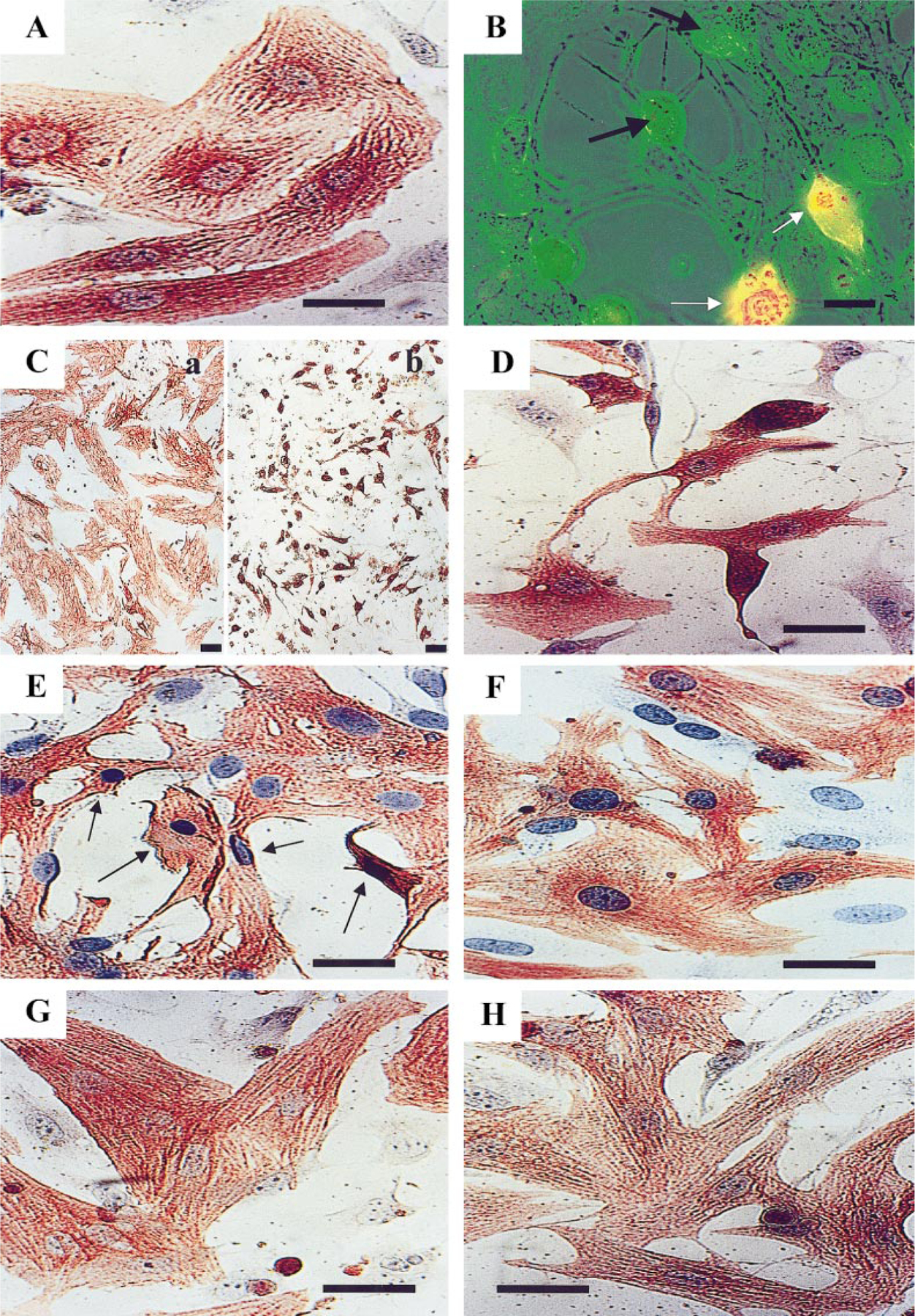
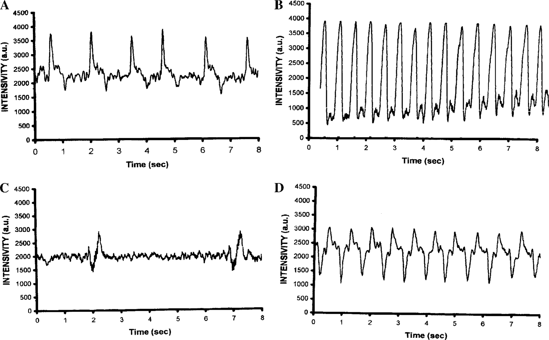
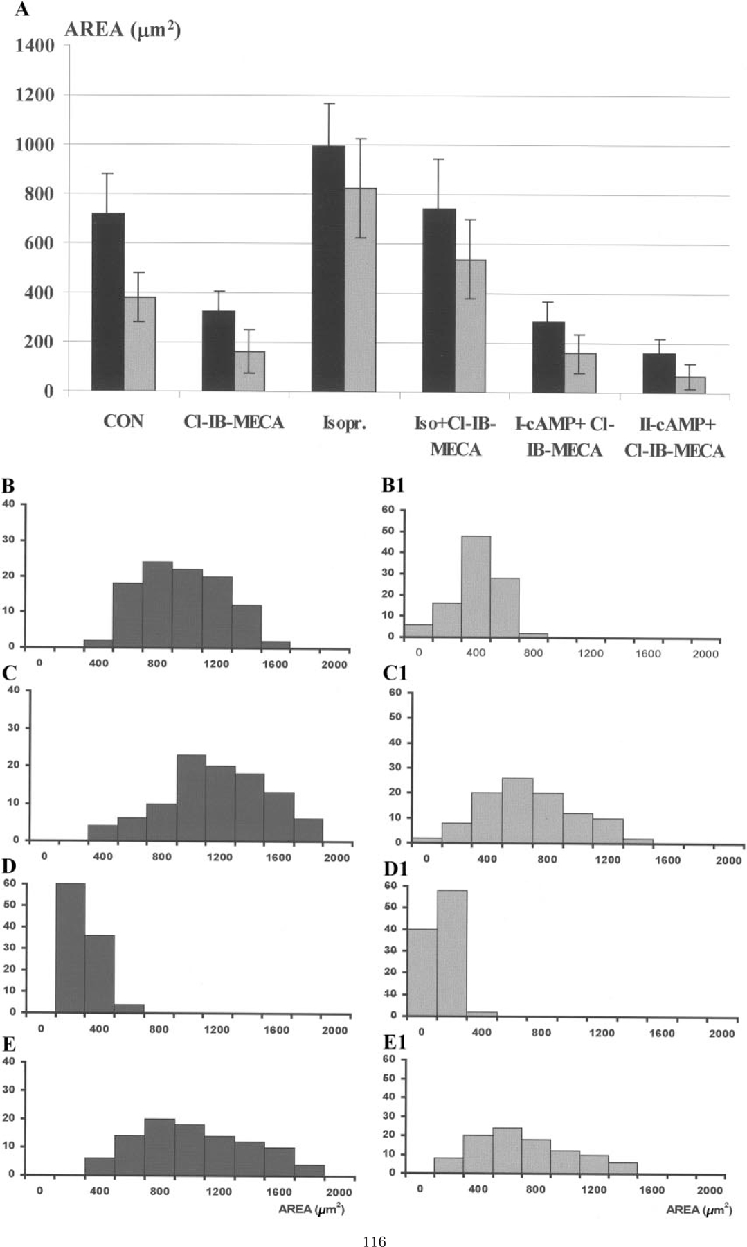
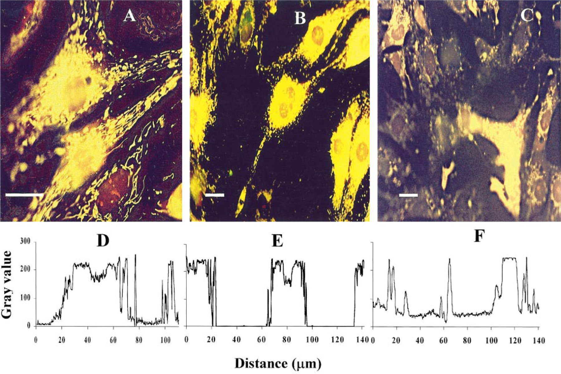
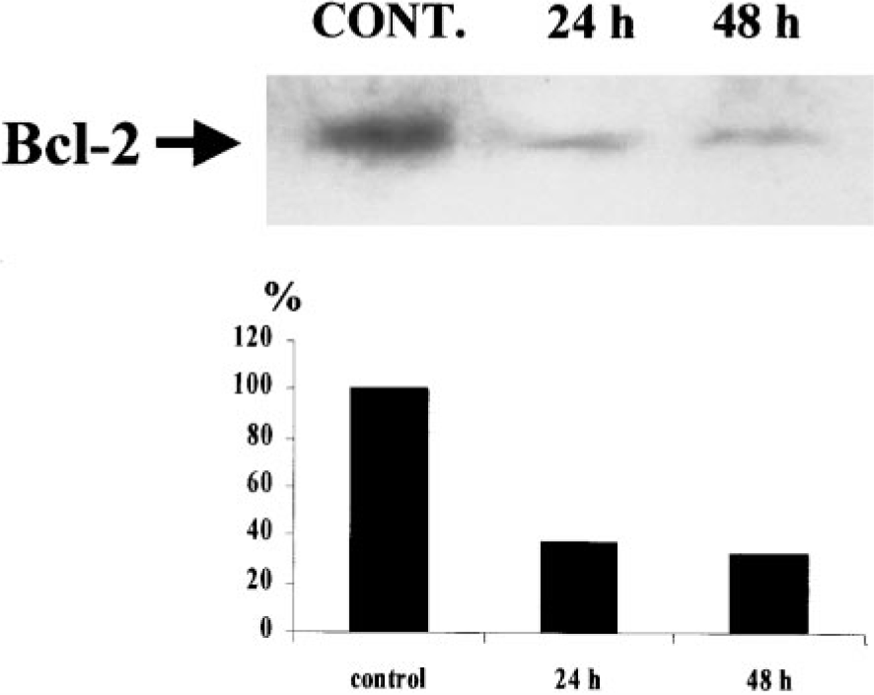
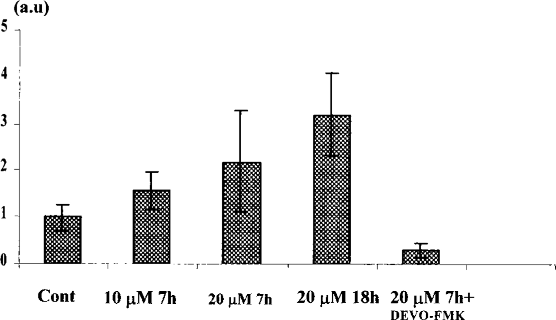
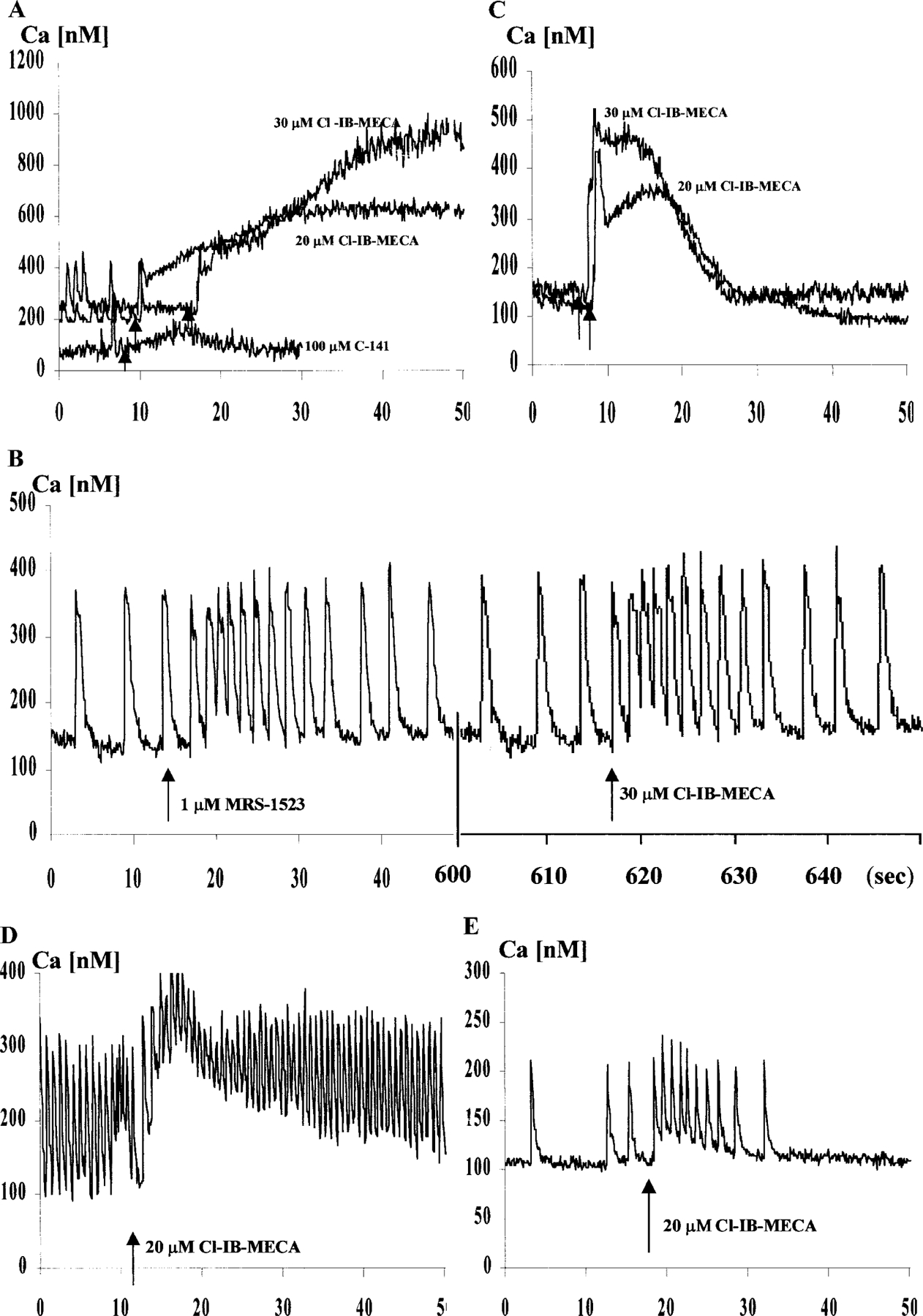
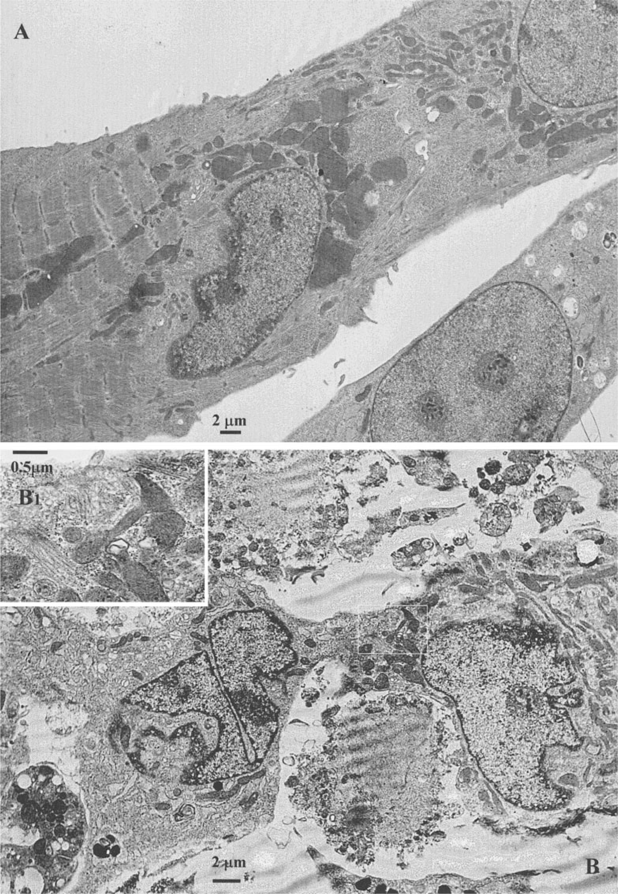
The purpose of the present study was to investigate the mechanisms involved in the induction of apoptosis in newborn cultured cardiomyocytes by activation of adenosine (ADO) A3 receptors and to examine the protective effects of beta-adrenoceptors. The selective agonist for A3 ADO receptors Cl-IB-MECA (2-chloro-N6-iodobenzyl-5-N-methylcarboxamidoadenosine) and the antagonist MRS1523 (5-propyl-2-ethyl-4-propyl-3-(ethylsulfanylcarbonyl)-6-phenylpy rid ine-5-carboxylate) were used. High concentrations of the Cl-IB-MECA (> or = 10 microM) agonist induced morphological modifications of myogenic cells, such as rounding and retraction of cell body and dissolution of contractile filaments, followed by apoptotic death. In addition, Cl-IB-MECA caused a sustained and reversible increase in [Ca2+]i, which was prevented by the selective antagonist MRS1523. Furthermore, MRS1523 protected the cardiocytes if briefly exposed to Cl-IB-MECA and partially protected from prolonged (48 h) agonist exposure. Apoptosis induced by Cl-IB-MECA was not redox-dependent, since the mitochondrial membrane potential remained constant until the terminal stage of cell death. Cl-IB-MECA activated caspase-3 protease in a concentration-dependent manner after 7 h of treatment and more effectively after 18 h of exposure. Bcl-2 protein was readily detected in control cells, and its expression was significantly decreased after 24 and 48 h of treatment with Cl-IB-MECA. Beta-adrenergic stimulation antagonized the pro-apoptotic effects of Cl-IB-MECA, probably through a cAMP/protein kinase A-independent mechanism, since addition of dibutyryl-cAMP did not abolish the apoptosis induced by Cl-IB-MECA. Incubation of cultured myocytes with isoproterenol (5 microM) for 3 or 24 h almost completely abolished the increase in [Ca2+]i. Prolonged incubation of cardiomyocytes with isoproterenol and Cl-IB-MECA did not induce apoptosis. Our data suggest that the apoptosis-inducing signal from activation of adenosine A3 receptors (or counteracting beta-adrenergic signal) leads to the activation of the G-protein-coupled enzymes and downstream pathways to a self-amplifying cascade. Expression of different genes within this cascade is responsible for orchestrating either cardiomyocyte apoptosis or its protection.
本研究的目的是探讨激活腺苷(ADO)A3受体诱导新生培养心肌细胞凋亡的机制,并研究β-肾上腺素能受体的保护作用。使用了A3 ADO受体的选择性激动剂Cl-IB-MECA(2-氯-N6-碘苄基-5-N-甲基甲酰胺腺苷)和拮抗剂MRS1523(5-丙基-2-乙基-4-丙基-3-(乙硫基羰基)-6-苯基吡啶-5-羧酸盐)。高浓度的Cl-IB-MECA(≥10μM)激动剂可诱导肌源性细胞发生形态学改变,如细胞体变圆、回缩以及收缩细丝溶解,随后发生凋亡死亡。此外,Cl-IB-MECA可使细胞内钙离子浓度([Ca2+]i)持续且可逆地升高,这一作用可被选择性拮抗剂MRS1523阻断。此外,如果短暂暴露于Cl-IB-MECA,MRS1523可保护心肌细胞,对于长时间(48小时)暴露于激动剂的情况也有部分保护作用。Cl-IB-MECA诱导的凋亡不依赖于氧化还原反应,因为线粒体膜电位在细胞死亡末期之前保持恒定。处理7小时后,Cl-IB-MECA以浓度依赖的方式激活caspase-3蛋白酶,暴露18小时后激活作用更有效。在对照细胞中可轻易检测到Bcl-2蛋白,用Cl-IB-MECA处理24小时和48小时后,其表达显著降低。β-肾上腺素能刺激拮抗Cl-IB-MECA的促凋亡作用,可能是通过一种不依赖于环磷酸腺苷/蛋白激酶A的机制,因为添加二丁酰环磷酸腺苷并不能消除Cl-IB-MECA诱导的凋亡。用异丙肾上腺素(5μM)孵育培养的心肌细胞3小时或24小时几乎完全消除了[Ca2+]i的升高。心肌细胞用异丙肾上腺素和Cl-IB-MECA长时间孵育不会诱导凋亡。我们的数据表明,腺苷A3受体激活(或对抗β-肾上腺素能信号)产生的凋亡诱导信号导致G蛋白偶联酶和下游通路激活,形成一个自我放大的级联反应。该级联反应中不同基因的表达负责协调心肌细胞凋亡或其保护过程。