Klingbeil C K, Hauck C R, Hsia D A, Jones K C, Reider S R, Schlaepfer D D
Department of Immunology, The Scripps Research Institute, La Jolla, California 92037, USA.
J Cell Biol. 2001 Jan 8;152(1):97-110. doi: 10.1083/jcb.152.1.97.
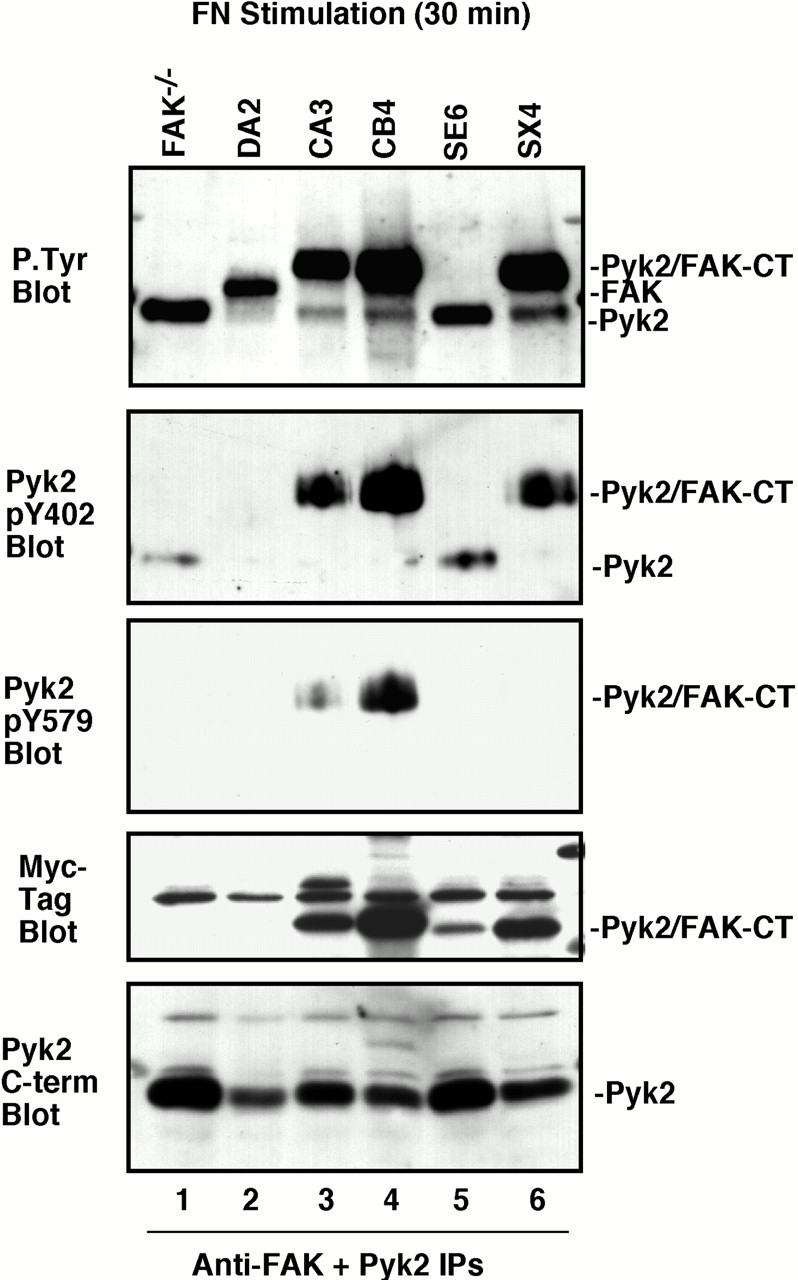
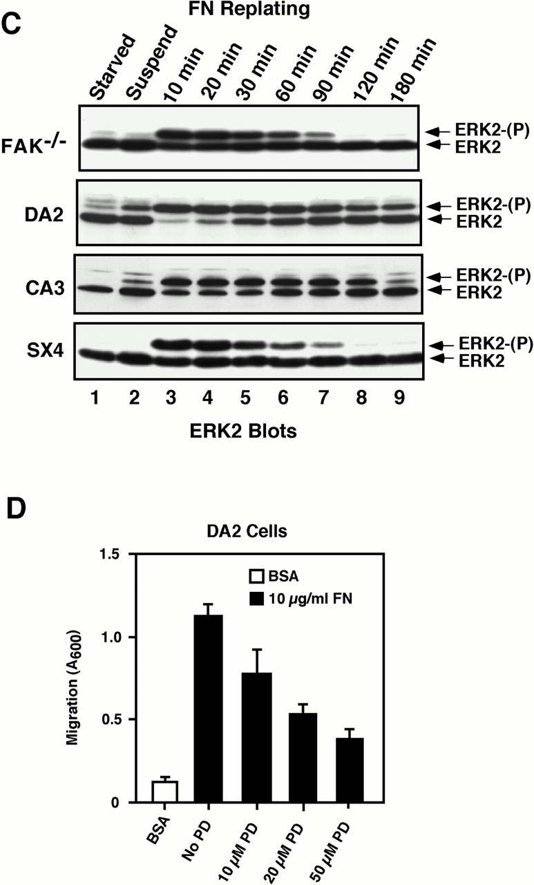
Focal adhesion kinase-null (FAK(-/-) fibroblasts exhibit morphological and motility defects that are reversed by focal adhesion kinase (FAK) reexpression. The FAK-related kinase, proline-rich tyrosine kinase 2 (Pyk2), is expressed in FAK(-/-) cells, yet it exhibits a perinuclear distribution and does not functionally substitute for FAK. Chimeric Pyk2/FAK proteins were created and expressed in FAK(-/-) cells to determine the impact of Pyk2 localization to focal contacts. Whereas an FAK/Pyk2 COOH-terminal (CT) domain chimera was perinuclear distributed, stable expression of a Pyk2 chimera with the FAK-CT domain (Pyk2/FAK-CT) localized to focal contact sites and enhanced fibronectin (FN)-stimulated haptotactic cell migration equal to FAK-reconstituted cells. Disruption of paxillin binding to the FAK-CT domain (S-1034) inhibited Pyk2/FAK-CT localization to focal contacts and its capacity to promote cell motility. Paxillin binding to the FAK-CT was necessary but not sufficient to mediate the indirect association of FAK or Pyk2/FAK-CT with a beta 1-integrin-containing complex. Both FAK and Pyk2/FAK-CT but not Pyk2/FAK-CT S-1034 reconstituted FAK(-/-) cells, exhibit elevated FN-stimulated extracellular signal-regulated kinase 2 (ERK2) and c-Jun NH(2)-terminal kinase (JNK) kinase activation. FN-stimulated FAK or Pyk2/FAK-CT activation enhanced both the extent and duration of FN-stimulated ERK2 activity which was necessary for cell motility. Transient overexpression of the FAK-CT but not FAK-CT S-1034 domain inhibited both FN-stimulated ERK2 and JNK activation as well as FN-stimulated motility of Pyk2/FAK-CT reconstituted cells. These gain-of-function studies show that the NH(2)-terminal and kinase domains of Pyk2 can functionally substitute for FAK in promoting FN-stimulated signaling and motility events when localized to beta-integrin-containing focal contact sites via interactions mediated by the FAK-CT domain.
粘着斑激酶缺失(FAK(-/-))的成纤维细胞表现出形态和运动缺陷,而粘着斑激酶(FAK)的重新表达可逆转这些缺陷。与FAK相关的激酶,富含脯氨酸的酪氨酸激酶2(Pyk2),在FAK(-/-)细胞中表达,但其呈核周分布,且在功能上不能替代FAK。构建了嵌合的Pyk2/FAK蛋白并在FAK(-/-)细胞中表达,以确定Pyk2定位于粘着斑的影响。虽然一个FAK/Pyk2羧基末端(CT)结构域嵌合体呈核周分布,但一个带有FAK-CT结构域的Pyk2嵌合体(Pyk2/FAK-CT)的稳定表达定位于粘着斑部位,并增强了纤连蛋白(FN)刺激的趋触性细胞迁移,其程度与FAK重构细胞相当。桩蛋白与FAK-CT结构域(S-1034)的结合破坏抑制了Pyk2/FAK-CT定位于粘着斑及其促进细胞运动的能力。桩蛋白与FAK-CT的结合对于介导FAK或Pyk2/FAK-CT与含β1整合素的复合物的间接结合是必要的,但并不充分。FAK和Pyk2/FAK-CT而非Pyk2/FAK-CT S-1034重构的FAK(-/-)细胞,表现出FN刺激的细胞外信号调节激酶2(ERK2)和c-Jun NH(2)-末端激酶(JNK)激酶活性升高。FN刺激的FAK或Pyk2/FAK-CT激活增强了FN刺激的ERK2活性的程度和持续时间,这对于细胞运动是必需的。FAK-CT结构域而非FAK-CT S-1034结构域的瞬时过表达抑制了FN刺激的ERK2和JNK激活以及Pyk2/FAK-CT重构细胞的FN刺激的运动。这些功能获得性研究表明,当通过FAK-CT结构域介导的相互作用定位于含β整合素的粘着斑部位时,Pyk2的氨基末端和激酶结构域在促进FN刺激的信号传导和运动事件中可在功能上替代FAK。