McCarthy A D, Etcheverry S B, Bruzzone L, Lettieri G, Barrio D A, Cortizo A M
Cátedra de Bioquímica Patológica and Division Química Analítica, Facultad de Ciencias Exactas, Universidad Nacional de La Plata, La Plata, Argentina.
BMC Cell Biol. 2001;2:16. doi: 10.1186/1471-2121-2-16. Epub 2001 Aug 2.
The tissue accumulation of protein-bound advanced glycation endproducts (AGE) may be involved in the etiology of diabetic chronic complications, including osteopenia. The aim of this study was to investigate the effect of an AGE-modified type I collagen substratum on the adhesion, spreading, proliferation and differentiation of rat osteosarcoma UMR106 and mouse non-transformed MC3T3E1 osteoblastic cells. We also studied the role of reactive oxygen species (ROS) and nitric oxide synthase (NOS) expression on these AGE-collagen mediated effects.
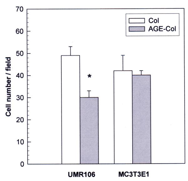
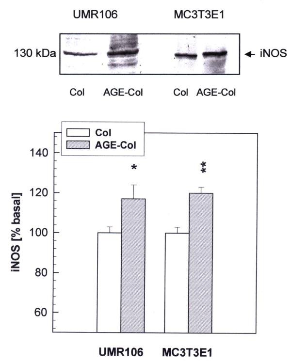
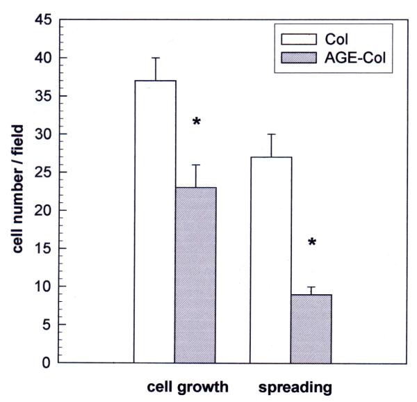
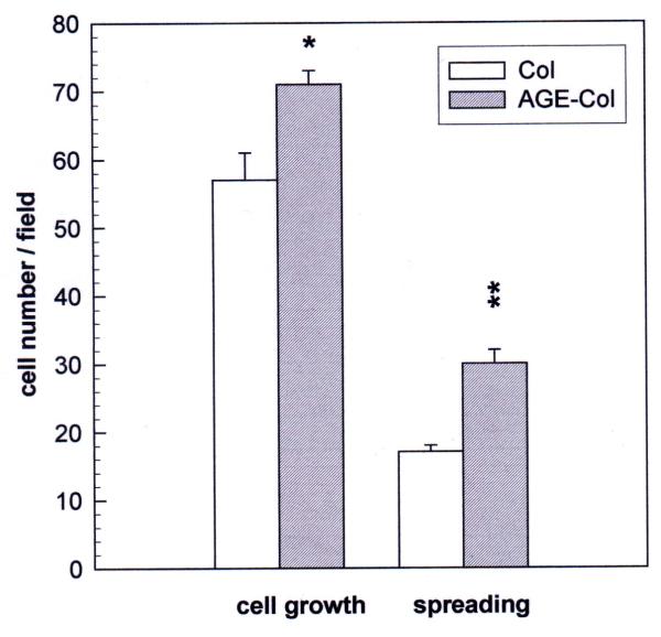
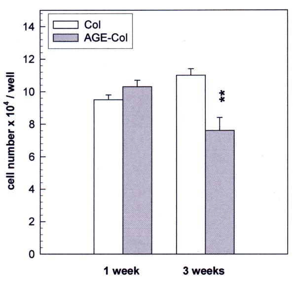
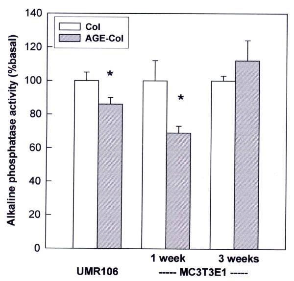
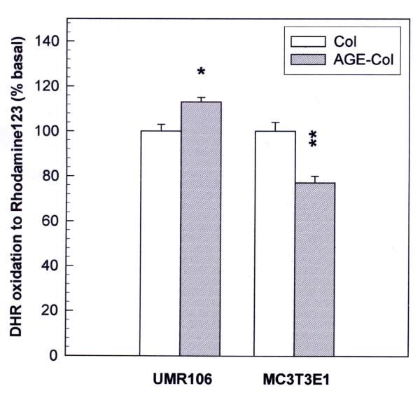
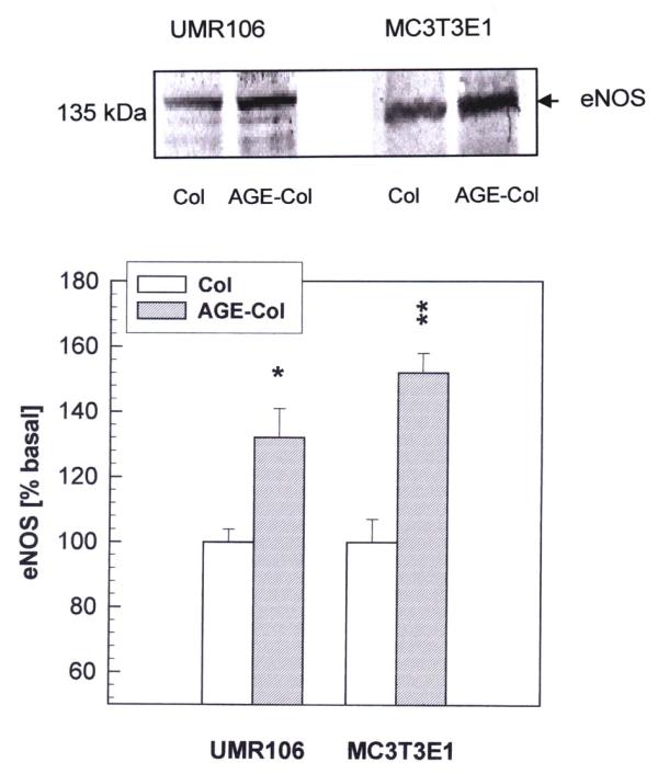
AGE-collagen decreased the adhesion of UMR106 cells, but had no effect on the attachment of MC3T3E1 cells. In the UMR106 cell line, AGE-collagen also inhibited cellular proliferation, spreading and alkaline phosphatase (ALP) activity. In preosteoblastic MC3T3E1 cells (24-hour culture), proliferation and spreading were significantly increased by AGE-collagen. After one week of culture (differentiated MC3T3E1 osteoblasts) AGE-collagen inhibited ALP activity, but had no effect on cell number. In mineralizing MC3T3E1 cells (3-week culture) AGE-collagen induced a decrease in the number of surviving cells and of extracellular nodules of mineralization, without modifying their ALP activity. Intracellular ROS production, measured after a 48-hour culture, was decreased by AGE-collagen in MC3T3E1 cells, but was increased by AGE-collagen in UMR106 cells. After a 24-hour culture, AGE-collagen increased the expression of endothelial and inducible NOS, in both osteoblastic cell lines.
These results suggest that the accumulation of AGE on bone extracellular matrix could regulate the proliferation and differentiation of osteoblastic cells. These effects appear to depend on the stage of osteoblastic development, and possibly involve the modulation of NOS expression and intracellular ROS pathways.
蛋白质结合的晚期糖基化终产物(AGE)在组织中的蓄积可能参与包括骨质减少在内的糖尿病慢性并发症的发病机制。本研究旨在探讨AGE修饰的I型胶原基质对大鼠骨肉瘤UMR106细胞和小鼠未转化的MC3T3E1成骨细胞的黏附、铺展、增殖和分化的影响。我们还研究了活性氧(ROS)和一氧化氮合酶(NOS)表达在这些AGE-胶原介导效应中的作用。
AGE-胶原降低了UMR106细胞的黏附,但对MC3T3E1细胞的附着没有影响。在UMR106细胞系中,AGE-胶原还抑制细胞增殖、铺展和碱性磷酸酶(ALP)活性。在成骨前MC3T3E1细胞(培养24小时)中,AGE-胶原显著增加了增殖和铺展。培养一周后(分化的MC3T3E1成骨细胞),AGE-胶原抑制了ALP活性,但对细胞数量没有影响。在矿化的MC3T3E1细胞(培养3周)中,AGE-胶原导致存活细胞数量和细胞外矿化结节减少,而不改变其ALP活性。培养48小时后测量的细胞内ROS产生,在MC3T3E1细胞中被AGE-胶原降低,但在UMR106细胞中被AGE-胶原增加。培养24小时后,AGE-胶原增加了两种成骨细胞系中内皮型和诱导型NOS的表达。
这些结果表明,AGE在骨细胞外基质上的蓄积可能调节成骨细胞的增殖和分化。这些效应似乎取决于成骨细胞发育阶段,并且可能涉及NOS表达和细胞内ROS途径的调节。