Ramsey M S, Dickson D H
Br J Ophthalmol. 1975 Jun;59(6):338-42. doi: 10.1136/bjo.59.6.338.
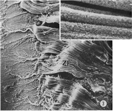
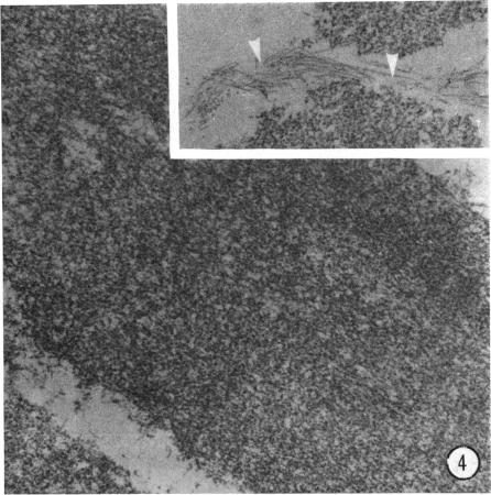
The lens from a patient with homocystinuria was examined by scanning and transmission electron microscopy. A fringe of zonular remnants was found attached to the anterior lens capsule, and was observed to be composed of masses of short filaments in disarray, together with occasional bundles of normal-appearing zonular filaments. Although a pericapsular membrane (zonular lamella) was not observed, the remainder of the lens capsule and epithelium appeared unremarkable. The lens fringe of white zonular remnants may be characteristic, if not pathognomic, for homocystinuria.
通过扫描电子显微镜和透射电子显微镜对一名同型胱氨酸尿症患者的晶状体进行了检查。发现有一圈小带残余物附着在前囊膜上,观察到其由杂乱的短纤维团块组成,偶尔还有外观正常的小带纤维束。虽然未观察到囊周膜(小带板层),但晶状体囊膜和上皮的其余部分看起来并无异常。白色小带残余物形成的晶状体边缘即便不是同型胱氨酸尿症的特异性表现,也可能是其特征性表现。