• 文献检索
  • 文档翻译
  • 深度研究
  • 学术资讯
  • Suppr Zotero 插件Zotero 插件
  • 邀请有礼
  • 套餐&价格
  • 历史记录
应用&插件
Suppr Zotero 插件Zotero 插件浏览器插件Mac 客户端Windows 客户端微信小程序
定价
高级版会员购买积分包购买API积分包
服务
文献检索文档翻译深度研究API 文档MCP 服务
关于我们
关于 Suppr公司介绍联系我们用户协议隐私条款
关注我们

Suppr 超能文献

核心技术专利:CN118964589B侵权必究
粤ICP备2023148730 号-1Suppr @ 2026

文献检索

告别复杂PubMed语法,用中文像聊天一样搜索,搜遍4000万医学文献。AI智能推荐,让科研检索更轻松。

立即免费搜索

文件翻译

保留排版,准确专业,支持PDF/Word/PPT等文件格式,支持 12+语言互译。

免费翻译文档

深度研究

AI帮你快速写综述,25分钟生成高质量综述,智能提取关键信息,辅助科研写作。

立即免费体验

乳腺结节的超声评估:传统、实时复合及脉冲反转谐波图像的比较

Sonographic evaluation of breast nodules: comparison of conventional, real-time compound, and pulse-inversion harmonic images.

作者信息

Seo Bo Kyoung, Oh Yu Whan, Kim Hyung Rae, Kim Hong Weon, Kang Chang Ho, Lee Nam Joon, Kim Jung Hyuk, Park Bum Jin, Cho Kyu Ran, Lee June Young, Lee Ki Yeoul, Bae Jeoung Won

机构信息

Department of Diagnostic Radiology, Korea University Anam Hospital, 126-1 Anam-dong 5-ga, Sungbuk-gu, Seoul 136-705, Korea.

出版信息

Korean J Radiol. 2002 Jan-Mar;3(1):38-44. doi: 10.3348/kjr.2002.3.1.38.

DOI:10.3348/kjr.2002.3.1.38
PMID:11919477
原文链接:https://pmc.ncbi.nlm.nih.gov/articles/PMC2713985/
Abstract

OBJECTIVE

To compare the use of conventional, real-time compound, and pulse-inversion harmonic imaging in the evaluation of breast nodules.

MATERIALS AND METHODS

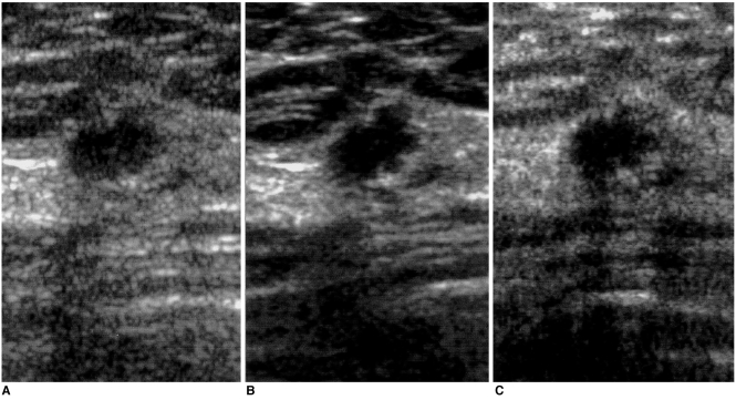
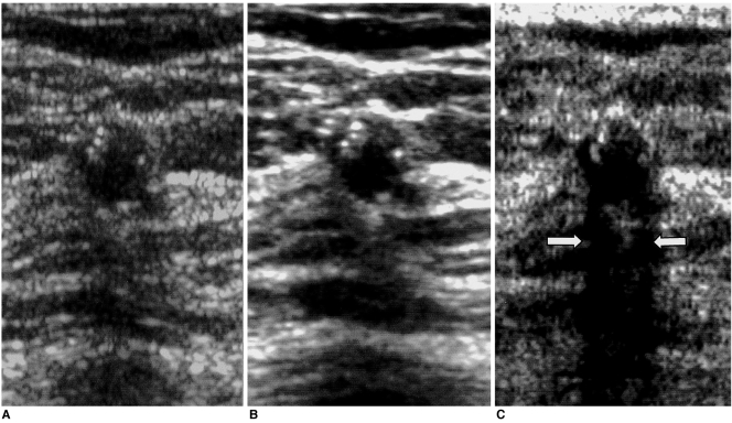
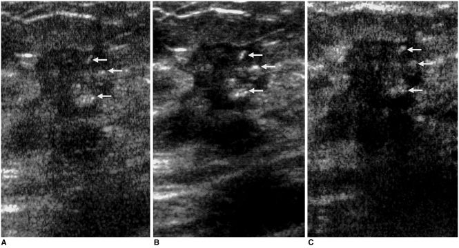
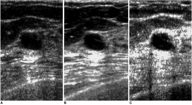
Fifty-two breast nodules were included in this study, conducted between May and December 2000, in which conventional, real-time compound, and pulse-inversion harmonic images were obtained in the same plane. Three radiologists, each blinded to the interpretations of the other two, evaluated the findings, characterizing the lesions and ranking the three techniques from grade 1, the worst, to grade 3, the best. Lesion conspicuity was assessed, and lesions were also characterized in terms of their margin, clarity of internal echotexture, and clarity of posterior echo pattern. The three techniques were compared using Friedman's test, and interobserver agreement in image interpretation was assessed by means of the intraclass correlation coefficient.

RESULTS

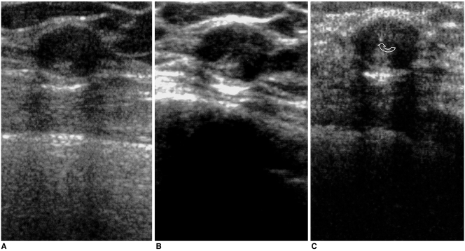
With regard to lesion conspicuity, margin, and internal echotexture of the nodules, real-time compound imaging was the best technique (p < 0.05); in terms of posterior echo pattern, the best was pulse-inversion harmonic imaging (p < 0.05). Real-time compound and pulse inversion harmonic imaging were better than conventional sonography in all evaluative aspects. Interobserver agreement was greater than moderate.

CONCLUSION

Real-time compound and pulse-inversion harmonic imaging procedures are superior to conventional sonography in terms of both lesion conspicuity and the further characterization of breast nodules. Real-time compound imaging is the best technique for evaluation of the margin and internal echotexture of nodules, while pulse-inversion harmonic imaging is very effective for the evaluation of the posterior echo patterns.

摘要

目的

比较传统成像、实时复合成像和脉冲反转谐波成像在乳腺结节评估中的应用。

材料与方法

本研究纳入了52个乳腺结节,研究于2000年5月至12月进行,在同一平面获取传统成像、实时复合成像和脉冲反转谐波图像。三名放射科医生分别对其他两人的解读不知情,对检查结果进行评估,对病变进行特征描述,并将这三种技术从最差的1级到最好的3级进行排序。评估病变的清晰度,还从病变边缘、内部回声纹理清晰度和后方回声模式清晰度方面对病变进行特征描述。使用Friedman检验比较这三种技术,并通过组内相关系数评估观察者间在图像解读方面的一致性。

结果

在结节的病变清晰度、边缘和内部回声纹理方面,实时复合成像为最佳技术(p<0.05);在后方回声模式方面,最佳的是脉冲反转谐波成像(p<0.05)。在所有评估方面,实时复合成像和脉冲反转谐波成像均优于传统超声检查。观察者间的一致性大于中等程度。

结论

在病变清晰度和乳腺结节的进一步特征描述方面,实时复合成像和脉冲反转谐波成像程序优于传统超声检查。实时复合成像是评估结节边缘和内部回声纹理的最佳技术,而脉冲反转谐波成像在评估后方回声模式方面非常有效。

https://cdn.ncbi.nlm.nih.gov/pmc/blobs/46ca/2713985/0b6abf04616a/kjr-3-38-g005.jpg
https://cdn.ncbi.nlm.nih.gov/pmc/blobs/46ca/2713985/47d3eff028b9/kjr-3-38-g001.jpg
https://cdn.ncbi.nlm.nih.gov/pmc/blobs/46ca/2713985/4dee7b436519/kjr-3-38-g002.jpg
https://cdn.ncbi.nlm.nih.gov/pmc/blobs/46ca/2713985/9991d9374dc1/kjr-3-38-g003.jpg
https://cdn.ncbi.nlm.nih.gov/pmc/blobs/46ca/2713985/066384199498/kjr-3-38-g004.jpg
https://cdn.ncbi.nlm.nih.gov/pmc/blobs/46ca/2713985/0b6abf04616a/kjr-3-38-g005.jpg

相似文献

1
Sonographic evaluation of breast nodules: comparison of conventional, real-time compound, and pulse-inversion harmonic images.乳腺结节的超声评估:传统、实时复合及脉冲反转谐波图像的比较
Korean J Radiol. 2002 Jan-Mar;3(1):38-44. doi: 10.3348/kjr.2002.3.1.38.
2
Ultrasonographic evaluation of focal hepatic lesions: comparison of pulse inversion harmonic, tissue harmonic, and conventional imaging techniques.肝脏局灶性病变的超声评估:脉冲反转谐波、组织谐波及传统成像技术的比较
J Ultrasound Med. 2000 May;19(5):293-9; quiz 301-2.
3
Tissue harmonic imaging, frequency compound imaging, and conventional imaging: use and benefit in breast sonography.组织谐波成像、频率复合成像与传统成像:在乳腺超声检查中的应用及优势
J Ultrasound Med. 2007 Aug;26(8):1041-51. doi: 10.7863/jum.2007.26.8.1041.
4
Comparison of conventional sonography, real-time compound sonography, tissue harmonic sonography, and tissue harmonic compound sonography of abdominal and pelvic lesions.腹部和盆腔病变的传统超声检查、实时复合超声检查、组织谐波超声检查及组织谐波复合超声检查的比较
AJR Am J Roentgenol. 2003 Nov;181(5):1341-7. doi: 10.2214/ajr.181.5.1811341.
5
Diagnostic accuracy of phase-inversion tissue harmonic imaging versus fundamental B-mode sonography in the evaluation of focal lesions of the kidney.在评估肾脏局灶性病变中,相控阵组织谐波成像与基波B型超声检查的诊断准确性比较
AJR Am J Roentgenol. 2003 Jun;180(6):1639-47. doi: 10.2214/ajr.180.6.1801639.
6
Contribution of tissue harmonic imaging and frequency compound imaging in interventional breast sonography.组织谐波成像和频率复合成像在乳腺介入超声检查中的作用
J Ultrasound Med. 2006 Jul;25(7):845-55. doi: 10.7863/jum.2006.25.7.845.
7
Characterization of benign and malignant solid breast masses: comparison of conventional US and tissue harmonic imaging.乳腺实性良恶性肿块的特征:传统超声与组织谐波成像的比较
Radiology. 2007 Jan;242(1):63-9. doi: 10.1148/radiol.2421050859. Epub 2006 Nov 7.
8
Comparing Levovist-enhanced pulse inversion harmonic imaging and ferumoxides-enhanced MR imaging of hepatic metastases.对比左旋糖酐铁增强脉冲反转谐波成像与超顺磁性氧化铁增强磁共振成像对肝转移瘤的诊断价值。
AJR Am J Roentgenol. 2003 May;180(5):1339-46. doi: 10.2214/ajr.180.5.1801339.
9
Comparison of conventional, compounding, computer enhancement, and compounding with computer enhancement in ultrasound imaging of the breast.传统超声成像、复合成像、计算机增强成像以及复合成像与计算机增强成像在乳腺超声成像中的比较。
Ultrasound Q. 2009 Sep;25(3):129-34. doi: 10.1097/RUQ.0b013e3181b739f1.
10
Hepatocellular carcinoma treated with radiofrequency ablation: comparison of pulse inversion contrast-enhanced harmonic sonography, contrast-enhanced power Doppler sonography, and helical CT.经射频消融治疗的肝细胞癌:脉冲反转对比增强谐波超声、对比增强功率多普勒超声及螺旋CT的比较
AJR Am J Roentgenol. 2001 Aug;177(2):375-80. doi: 10.2214/ajr.177.2.1770375.

引用本文的文献

1
Ultrasound as a Method for Early Diagnosis of Breast Pathology.超声作为乳腺疾病早期诊断的一种方法。
J Pers Med. 2023 Jul 18;13(7):1156. doi: 10.3390/jpm13071156.
2
A new method for assessing the performance of signal processing filters in suppressing the side lobe level.一种评估信号处理滤波器抑制旁瓣电平性能的新方法。
Ultrasonography. 2021 Apr;40(2):289-300. doi: 10.14366/usg.20032. Epub 2020 Jul 15.
3
Comparison of conventional B-scan, tissue harmonic imaging, compound imaging and tissue harmonic compound imaging in neck lesion characterisation.

本文引用的文献

1
Intraclass correlations: uses in assessing rater reliability.组内相关系数:在评估评分者可靠性中的应用。
Psychol Bull. 1979 Mar;86(2):420-8. doi: 10.1037//0033-2909.86.2.420.
2
Detection of parenchymal abnormalities in acute pyelonephritis by pulse inversion harmonic imaging with or without microbubble ultrasonographic contrast agent: correlation with computed tomography.使用或不使用微泡超声造影剂的脉冲反转谐波成像检测急性肾盂肾炎实质异常:与计算机断层扫描的相关性
J Ultrasound Med. 2001 Jan;20(1):5-14. doi: 10.7863/jum.2001.20.1.5.
3
Ultrasonographic evaluation of focal hepatic lesions: comparison of pulse inversion harmonic, tissue harmonic, and conventional imaging techniques.
比较常规 B 超、组织谐波成像、复合成像和组织谐波复合成像在颈部病变特征中的应用。
Eur Arch Otorhinolaryngol. 2010 Oct;267(10):1593-8. doi: 10.1007/s00405-010-1255-6. Epub 2010 Apr 27.
4
Comparative analysis of logistic regression, support vector machine and artificial neural network for the differential diagnosis of benign and malignant solid breast tumors by the use of three-dimensional power Doppler imaging.利用三维能量多普勒成像对乳腺实性良恶性肿瘤进行鉴别诊断时,逻辑回归、支持向量机和人工神经网络的比较分析
Korean J Radiol. 2009 Sep-Oct;10(5):464-71. doi: 10.3348/kjr.2009.10.5.464. Epub 2009 Aug 25.
5
Value of tissue harmonic imaging (THI) and contrast harmonic imaging (CHI) in detection and characterisation of breast tumours.组织谐波成像(THI)和对比谐波成像(CHI)在乳腺肿瘤检测与特征描述中的价值。
Eur Radiol. 2007 Jan;17(1):1-10. doi: 10.1007/s00330-006-0325-z. Epub 2006 Jul 6.
6
Comparison of fundamental sonography, tissue-harmonic sonography, fundamental compound sonography, and tissue-harmonic compound sonography for focal hepatic lesions.基础超声、组织谐波超声、基础复合超声及组织谐波复合超声对肝脏局灶性病变的比较
Eur Radiol. 2006 Nov;16(11):2444-53. doi: 10.1007/s00330-006-0205-6. Epub 2006 Mar 29.
7
Phase-inversion tissue harmonic imaging compared to fundamental B-mode ultrasound in the evaluation of the pathology of large and small bowel.与基波B型超声相比,相位反转组织谐波成像在评估大肠和小肠病理中的应用
Eur Radiol. 2005 Sep;15(9):2021-30. doi: 10.1007/s00330-005-2749-2. Epub 2005 Apr 8.
肝脏局灶性病变的超声评估:脉冲反转谐波、组织谐波及传统成像技术的比较
J Ultrasound Med. 2000 May;19(5):293-9; quiz 301-2.
4
Comparison of harmonic and conventional power Doppler ultrasonography for assessment of slow flow in hyperechoic tissue: experimental study using a Doppler phantom.谐波与传统能量多普勒超声评估高回声组织中缓慢血流的比较:使用多普勒体模的实验研究
Invest Radiol. 2000 Feb;35(2):105-10. doi: 10.1097/00004424-200002000-00003.
5
Pulse inversion imaging of liver blood flow: improved method for characterizing focal masses with microbubble contrast.肝脏血流的脉冲反转成像:利用微泡造影剂表征局灶性肿块的改进方法
Invest Radiol. 2000 Jan;35(1):58-71. doi: 10.1097/00004424-200001000-00007.
6
Tissue harmonic imaging sonography: evaluation of image quality compared with conventional sonography.组织谐波成像超声检查:与传统超声检查相比的图像质量评估
AJR Am J Roentgenol. 1998 Nov;171(5):1203-6. doi: 10.2214/ajr.171.5.9798848.
7
Multi-angle compound imaging.
Ultrason Imaging. 1998 Apr;20(2):81-102. doi: 10.1177/016173469802000201.
8
Breast imaging and the standard of care for the symptomatic patient.
Radiology. 1993 Jun;187(3):608-11. doi: 10.1148/radiology.187.3.8497602.
9
Solid breast nodules: use of sonography to distinguish between benign and malignant lesions.实性乳腺结节:利用超声鉴别良恶性病变
Radiology. 1995 Jul;196(1):123-34. doi: 10.1148/radiology.196.1.7784555.
10
A multimode real time scanner.一种多模式实时扫描仪。
Ultrasound Med Biol. 1980;6(3):279-84. doi: 10.1016/0301-5629(80)90024-1.