Mosesson M W, Amrani D L, Ménaché D
J Clin Invest. 1976 Mar;57(3):782-90. doi: 10.1172/JCI108337.
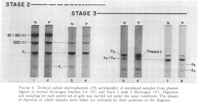
The structural properties of an inherited fibrinogen abnormality designated fibrinogen Paris I were investigated. Dodecyl sulfate gel electrophoresis of unreduced samples revealed no discernible differences in molecular weight from normal; this implied that in fibrinogen Paris I, the normal fibrinogen architecture of six covalently linked chains per molecule is preserved. Examination of dithiothreitol reduced samples before and after treatment with Reptilase or thrombin revealed that the Aalpha- and Bbeta-chains could release the A and B peptides, respectively. A mutant chain (mol wt 52,500, termed gammaParis I) which replaces a large proportion of gamma-chains (mol wt 49,400) was shown, like normal gamma-chains, to lack thrombin- and Reptilase-sensitive sites. The gamma-chains and alpha-chains of Paris I fibrin underwent Factor XIIIa-catalyzed cross-linking slowly; this behavior was not attributable to an intrinsic abnormality of these chains themselves but rather to the inhibitory effect of the mutant gammaParis I chains on this process. Results of DEAE-cellulose gradient elution chromatography of Paris I fibrinogen preparations revealed the presence of small amounts of normal fibrinogen molecules and also indicated that the gammaParis I chains possessed structural overlap with gamma-chains. Unlike gamma-chains however, the gammaParis I chains did not incorporate dansylcadaverine in the prescence of Factor XIIIa, nor, as previously reported, did they undergo cross-linking. The observations indicate that the amine acceptor site found in the COOH-terminal region of the gamma-chain is either not present on the gammaParis I chain or is unavailable for cross-linking. Further support for localization of the abnormality in the COOH-terminal region of the molecule was obtained from the observation that during plasmic hydrolysis of Paris I fibrinogen, at least one unique form of core Fragment D (DParis I) was evolved, whereas Fragment E did not differ from normal.
对一种名为纤维蛋白原巴黎I型的遗传性纤维蛋白原异常的结构特性进行了研究。未还原样品的十二烷基硫酸钠凝胶电泳显示,其分子量与正常情况无明显差异;这表明在纤维蛋白原巴黎I型中,每个分子由六条共价连接链组成的正常纤维蛋白原结构得以保留。对用二硫苏糖醇还原后的样品在使用蛇毒凝血酶或凝血酶处理前后进行检查发现,Aα链和Bβ链可分别释放A肽和B肽。已显示,一种取代了大部分γ链(分子量49,400)的突变链(分子量52,500,称为γ巴黎I型),与正常γ链一样,缺乏凝血酶和蛇毒凝血酶敏感位点。巴黎I型纤维蛋白的γ链和α链在因子XIIIa催化下的交联反应缓慢;这种行为并非归因于这些链本身的内在异常,而是由于突变的γ巴黎I型链对这一过程的抑制作用。巴黎I型纤维蛋白原制剂的DEAE - 纤维素梯度洗脱色谱结果显示存在少量正常纤维蛋白原分子,也表明γ巴黎I型链与γ链存在结构重叠。然而,与γ链不同的是,γ巴黎I型链在因子XIIIa存在的情况下不掺入丹磺酰尸胺,也不像先前报道的那样发生交联。这些观察结果表明,γ链COOH末端区域中发现的胺受体位点在γ巴黎I型链上要么不存在,要么无法用于交联。从巴黎I型纤维蛋白原的血浆水解过程中观察到至少一种独特形式的核心片段D(D巴黎I型)形成,而片段E与正常情况无异,这一现象进一步支持了异常位于分子COOH末端区域的观点。