• 文献检索
  • 文档翻译
  • 深度研究
  • 学术资讯
  • Suppr Zotero 插件Zotero 插件
  • 邀请有礼
  • 套餐&价格
  • 历史记录
应用&插件
Suppr Zotero 插件Zotero 插件浏览器插件Mac 客户端Windows 客户端微信小程序
定价
高级版会员购买积分包购买API积分包
服务
文献检索文档翻译深度研究API 文档MCP 服务
关于我们
关于 Suppr公司介绍联系我们用户协议隐私条款
关注我们

Suppr 超能文献

核心技术专利:CN118964589B侵权必究
粤ICP备2023148730 号-1Suppr @ 2026

文献检索

告别复杂PubMed语法,用中文像聊天一样搜索,搜遍4000万医学文献。AI智能推荐,让科研检索更轻松。

立即免费搜索

文件翻译

保留排版,准确专业,支持PDF/Word/PPT等文件格式,支持 12+语言互译。

免费翻译文档

深度研究

AI帮你快速写综述,25分钟生成高质量综述,智能提取关键信息,辅助科研写作。

立即免费体验

A quantitative assessment of the structural changes the rat's liver following obstruction of the common bile duct.

作者信息

Johnstone J M, Lee E G

出版信息

Br J Exp Pathol. 1976 Feb;57(1):85-94.

PMID:1268043
原文链接:https://pmc.ncbi.nlm.nih.gov/articles/PMC2041182/
Abstract

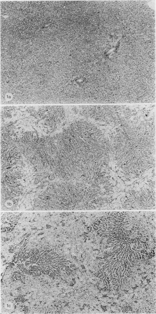
A study has been made of sequential changes in the rat's liver from 1 to 40 days after obstruction of the common bile duct. The qualitative changes have been described and illustrated. The volume proportions of hepatocytes, bile duct epithelium and biliary stroma have been quantified by histological analysis using a point counting technique. The proliferation of hepatocytes and bile duct cells have been measured by labelling with tritiated thymidine. The absolute quantity of hepatocytes in each liver has been estimated and expressed as a percentage of body weight. Over 40 days there is a relative fall in the volume proportion of hepatocytes and an increase in bile duct cells and biliary stroma. These changes in volume proportions are related directly to the period of jaundice. Biliary stroma increases in support of new bile duct tissue and there is no excessive fibrosis. Hepatocytes proliferate at a greater rate than normal after obstruction of the common bile duct and the degree of proliferation reaches a maximum of 24 times that of normal 4 days after obstruction. Similarly, the proliferation of bile duct epithelium is increased in obstructive jaundice but in this instance it reaches a maximum of 50 times that of normal 24 h after ligation of the common bile duct. The absolute quantity of hepatocytes in the liver probably falls during the period of jaundice. However, the fall is less than anticipated from the volume proportion of hepatocytes because of the overall increase in liver size.

摘要
https://cdn.ncbi.nlm.nih.gov/pmc/blobs/3826/2041182/7f350f6715e6/brjexppathol00139-0101-c.jpg
https://cdn.ncbi.nlm.nih.gov/pmc/blobs/3826/2041182/0a5bc240058f/brjexppathol00139-0100-a.jpg
https://cdn.ncbi.nlm.nih.gov/pmc/blobs/3826/2041182/0b68fbb9f688/brjexppathol00139-0101-a.jpg
https://cdn.ncbi.nlm.nih.gov/pmc/blobs/3826/2041182/5005621fb210/brjexppathol00139-0101-b.jpg
https://cdn.ncbi.nlm.nih.gov/pmc/blobs/3826/2041182/7f350f6715e6/brjexppathol00139-0101-c.jpg

相似文献

1
A quantitative assessment of the structural changes the rat's liver following obstruction of the common bile duct.
Br J Exp Pathol. 1976 Feb;57(1):85-94.
2
Proliferation and phenotypic modulation of portal fibroblasts in the early stages of cholestatic fibrosis in the rat.大鼠胆汁淤积性肝纤维化早期门静脉成纤维细胞的增殖及表型调节
Lab Invest. 1996 Jan;74(1):265-78.
3
The effect of ischemia on the regeneration of the cholestatic liver. An experimental study.缺血对胆汁淤积性肝再生的影响。一项实验研究。
Hepatogastroenterology. 2002 Mar-Apr;49(44):456-60.
4
Factors producing bile infarction and bile duct proliferation in biliary obstruction.导致胆道梗阻时胆汁梗死和胆管增生的因素。
J Pathol. 1990 Jan;160(1):57-62. doi: 10.1002/path.1711600112.
5
Somatostatin analogue (octreotide) inhibits bile duct epithelial cell proliferation and fibrosis after extrahepatic biliary obstruction.生长抑素类似物(奥曲肽)可抑制肝外胆管梗阻后胆管上皮细胞增殖及纤维化。
Am J Pathol. 1993 Dec;143(6):1574-8.
6
Histogenesis of bile duct-like cells proliferating during ethionine hepatocarcinogenesis. Evidence for a biliary epithelial nature of oval cells.乙硫氨酸诱导肝癌发生过程中增殖的胆管样细胞的组织发生。卵圆细胞具有胆管上皮性质的证据。
Lab Invest. 1992 Mar;66(3):390-402.
7
Alterations in bile ducts and peribiliary microcirculation in rats after common bile duct ligation.胆总管结扎后大鼠胆管及胆管周围微循环的改变
Hepatology. 1995 May;21(5):1380-6.
8
Serum alpha-fetoprotein in rats after ligation of the common bile duct: relation to ductular cell (oval cell) proliferation.胆总管结扎后大鼠血清甲胎蛋白:与小胆管细胞(卵圆细胞)增殖的关系
J Pathol. 1981 Jan;133(1):61-74. doi: 10.1002/path.1711330107.
9
Vitamin A deficiency associated with enhanced proliferation of bile duct epithelial cells in the rat.维生素A缺乏与大鼠胆管上皮细胞增殖增强有关。
Isr Med Assoc J. 2010 Feb;12(2):82-6.
10
The histoautoradiographic localization of taurocholate in rat liver after bile duct ligation. Evidence for ongoing secretion and reabsorption processes.胆管结扎后大鼠肝脏中牛磺胆酸盐的组织放射自显影定位。持续分泌和重吸收过程的证据。
J Hepatol. 1989 Mar;8(2):181-91. doi: 10.1016/0168-8278(89)90006-8.

引用本文的文献

1
Determination of 25-Hydroxyvitamin D in Rat Brain by Derivatization-Assisted LC/ESI-MS/MS.衍生化辅助液相色谱/电喷雾串联质谱法测定大鼠脑中的25-羟基维生素D
Biomed Chromatogr. 2025 Jul;39(7):e70119. doi: 10.1002/bmc.70119.
2
and Studies of the Hepatoprotective Activity of -Butylhydroquinone.关于 -丁基对苯二酚的肝保护活性的研究。
Int J Mol Sci. 2023 Dec 29;25(1):475. doi: 10.3390/ijms25010475.
3
A modified animal model of hepatic regeneration induced by hilar bile duct ligation.一种改良的肝再生动物模型,通过肝门胆管结扎诱导。

本文引用的文献

1
HEPATIC ALTERATIONS IN OBSTRUCTIVE JAUNDICE.阻塞性黄疸中的肝脏改变
Am J Clin Pathol. 1964 Feb;41:126-39. doi: 10.1093/ajcp/41.2.126.
2
The ductular cell reaction of rat liver in extrahepatic cholestasis. I. Proliferated biliary epithelial cells.大鼠肝在肝外胆汁淤积时的小胆管细胞反应。I. 增生的胆管上皮细胞。
Exp Mol Pathol. 1962 Apr;1:162-85. doi: 10.1016/0014-4800(62)90019-9.
3
Disturbances of structure and function in the liver as the result of biliary obstruction.胆管阻塞导致的肝脏结构和功能紊乱。
Sci Rep. 2021 Oct 12;11(1):20201. doi: 10.1038/s41598-021-99758-z.
4
Activation of WNT/Beta-Catenin Signaling and Regulation of the Farnesoid X Receptor/Beta-Catenin Complex After Murine Bile Duct Ligation.小鼠胆管结扎后WNT/β-连环蛋白信号通路的激活及法尼酯X受体/β-连环蛋白复合物的调控
Hepatol Commun. 2019 Oct 14;3(12):1642-1655. doi: 10.1002/hep4.1430. eCollection 2019 Dec.
5
Induction of chronic cholestasis without liver cirrhosis - Creation of an animal model.诱导非肝硬化性慢性胆汁淤积-动物模型的建立。
World J Gastroenterol. 2017 Jun 21;23(23):4191-4199. doi: 10.3748/wjg.v23.i23.4191.
6
Black blood T1rho MR imaging may diagnose early stage liver fibrosis: a proof-of-principle study with rat biliary duct ligation model.黑血T1rho磁共振成像可诊断早期肝纤维化:大鼠胆管结扎模型的原理验证研究
Quant Imaging Med Surg. 2016 Aug;6(4):353-363. doi: 10.21037/qims.2016.08.11.
7
Bone marrow-derived mesenchymal stem cells effectively regenerate fibrotic liver in bile duct ligation rat model.骨髓间充质干细胞可有效再生胆管结扎大鼠模型中的纤维化肝脏。
Exp Biol Med (Maywood). 2016 Mar;241(6):581-91. doi: 10.1177/1535370215627219. Epub 2016 Jan 24.
8
Animal Models for Fibrotic Liver Diseases: What We Have, What We Need, and What Is under Development.动物纤维化肝脏疾病模型:我们拥有的、需要的和正在开发的。
J Clin Transl Hepatol. 2015 Mar;3(1):53-66. doi: 10.14218/JCTH.2014.00035. Epub 2015 Mar 15.
9
Preexisting epithelial diversity in normal human livers: a tissue-tethered cytometric analysis in portal/periportal epithelial cells.正常人类肝脏中预先存在的上皮细胞多样性:门脉/周围上皮细胞中的组织束缚细胞计量分析。
Hepatology. 2013 Apr;57(4):1632-43. doi: 10.1002/hep.26131. Epub 2013 Mar 19.
10
Time course of collagen peak in bile duct-ligated rats.胆管结扎大鼠胆汁中胶原峰值的时间进程。
BMC Gastroenterol. 2011 Apr 28;11:45. doi: 10.1186/1471-230X-11-45.
J Pathol Bacteriol. 1958 Apr;75(2):333-49. doi: 10.1002/path.1700750212.
4
The effect of experimental bile-duct obstruction on critical biosynthetic functions of the liver.
Br J Surg. 1972 Jul;59(7):564-8. doi: 10.1002/bjs.1800590716.
5
The effect of obstructive jaundice on the migration of reticulo-endothelial cells and fibroblasts into early experimental granulomata.
Br J Surg. 1972 Nov;59(11):875-7. doi: 10.1002/bjs.1800591107.