McDermott Alison M, Redfern Rachel L, Zhang Bei, Pei Ying, Huang Ling, Proske Rita J
College of Optometry, University of Houston, Houston, Texas 77204-2020, USA.
Invest Ophthalmol Vis Sci. 2003 May;44(5):1859-65. doi: 10.1167/iovs.02-0787.
To investigate the expression of human beta-defensins (hBDs) by human corneal epithelium and determine the effects of proinflammatory cytokines on expression of human beta-defensin (hBD)-2 by human corneal epithelial cells (HCECs) in culture.
RNA was extracted from corneal epithelial cells scraped from cadaveric corneas and from cultured HCECs, and RT-PCR was performed to detect hBD-1, -2, and -3 mRNA. To study the effects of proinflammatory cytokines on expression of defensin, HCECs were cultured and then exposed to interleukin (IL)-1beta or tumor necrosis factor (TNF)-alpha for up to 36 hours, with a range of concentrations (0.01-100 ng/mL). In some experiments, cells were pretreated with various cell signaling pathway inhibitors before the addition of IL-1beta. At the end of the incubations, the cells were harvested for RT-PCR and the culture media collected for the detection by immunoblot analysis of secreted defensin peptide.
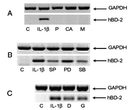
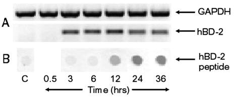
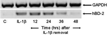
All epithelial tissue collected from cadaveric corneas expressed mRNA for hBD-1. hBD-2 was detectable in two of eight donors corneas, whereas hBD-3 was detected in five. All primary cultures of HCECs expressed hBD-1 and -3. A faint band for hBD-2 was detectable in three of eight cultures. Cultures of simian virus (SV)40-transformed HCECs always expressed hBD-1 and -3, but did not express hBD-2 under control conditions. IL-1beta and TNFalpha each stimulated the expression of hBD-2 in HCECs and were more effective in combination than alone. The effects of IL-1beta were concentration- (maximal at 10 ng/mL) and time-dependent (maximal at 12 hours and 24 hours for hBD-2 mRNA expression and protein secretion, respectively). The upregulation of hBD-2 mRNA persisted for at least 24 hours after removal of IL-1beta. The NFkappaB inhibitors pyrrolidinedithiocarbamate (PDTC; 100 microM), caffeic acid phenethyl ester (CAPE; 90 microM), and MG-132 (25 microM), blocked IL-1beta-stimulated expression of hBD-2. The p38 mitogen-activated protein (MAP) kinase inhibitor SB203580 (5 microM) and the c-Jun NH2-terminal kinase (JNK) inhibitor SP600125 (25 microM) partially blocked (by 47% and 59%, respectively) the effect of IL-1beta. However, PD98059, an ERK inhibitor, had no effect. Genistein (50 microM) and dexamethasone (1 microM) also partially blocked (by 26% and 28%, respectively) the effect of IL-1beta.
Human corneal epithelium expresses hBD-1 and -3. hBD-2 is not typically present, but its expression can be stimulated by proinflammatory cytokines such as IL-1beta, acting through mitogen-activated protein (MAP) kinase and nuclear factor (NF)-kappaB pathways. Because IL-1 is known to be increased at the ocular surface after injury, the current observations provide a mechanism to explain the previous finding that hBD-2 is upregulated in regenerating corneal epithelium. Cytokine stimulation of hBD-2 expression most likely provides additional protection against infection and raises the possibility that this defensin in particular may be involved in the wound-healing response, per se.

研究人角膜上皮细胞中人类β-防御素(hBDs)的表达,并确定促炎细胞因子对培养的人角膜上皮细胞(HCECs)中人类β-防御素(hBD)-2表达的影响。
从尸体角膜刮取的角膜上皮细胞和培养的HCECs中提取RNA,进行逆转录聚合酶链反应(RT-PCR)以检测hBD-1、-2和-3 mRNA。为研究促炎细胞因子对防御素表达的影响,培养HCECs,然后将其暴露于白细胞介素(IL)-1β或肿瘤坏死因子(TNF)-α长达36小时,浓度范围为0.01 - 100 ng/mL。在一些实验中,在添加IL-1β之前,用各种细胞信号通路抑制剂预处理细胞。孵育结束时,收获细胞进行RT-PCR,并收集培养基通过免疫印迹分析检测分泌的防御素肽。
从尸体角膜收集的所有上皮组织均表达hBD-1 mRNA。在8个供体角膜中的2个中可检测到hBD-2,而在5个中检测到hBD-3。所有HCECs的原代培养物均表达hBD-1和-3。在8个培养物中的3个中可检测到hBD-2的微弱条带。猿猴病毒(SV)40转化的HCECs培养物总是表达hBD-1和-3,但在对照条件下不表达hBD-2。IL-1β和TNFα均刺激HCECs中hBD-2的表达,且联合使用比单独使用更有效。IL-1β的作用具有浓度依赖性(在10 ng/mL时最大)和时间依赖性(hBD-2 mRNA表达和蛋白质分泌分别在12小时和24小时时最大)。去除IL-1β后,hBD-2 mRNA的上调持续至少24小时。NFκB抑制剂吡咯烷二硫代氨基甲酸盐(PDTC;100 μM)、咖啡酸苯乙酯(CAPE;90 μM)和MG-132(25 μM)可阻断IL-1β刺激的hBD-2表达。p38丝裂原活化蛋白(MAP)激酶抑制剂SB203580(5 μM)和c-Jun NH2末端激酶(JNK)抑制剂SP600125(25 μM)部分阻断(分别为47%和59%)IL-1β的作用。然而,ERK抑制剂PD98059没有作用。染料木黄酮(50 μM)和地塞米松(1 μM)也部分阻断(分别为26%和28%)IL-1β的作用。
人角膜上皮表达hBD-1和-3。hBD-2通常不存在,但其表达可被促炎细胞因子如IL-1β通过丝裂原活化蛋白(MAP)激酶和核因子(NF)-κB途径刺激。因为已知损伤后眼表IL-1会增加,目前的观察结果提供了一种机制来解释先前的发现,即hBD-2在再生角膜上皮中上调。细胞因子对hBD-2表达的刺激很可能提供额外的抗感染保护,并增加了这种防御素本身可能参与伤口愈合反应的可能性。