Jeyaraj D Antony, Grossman Gail, Petrusz Peter
Department of Cell and Developmental Biology and Laboratories for Reproductive Biology, University of North Carolina, Chapel Hill, NC 27599, USA.
Reprod Biol Endocrinol. 2003 Jun 12;1:48. doi: 10.1186/1477-7827-1-48.
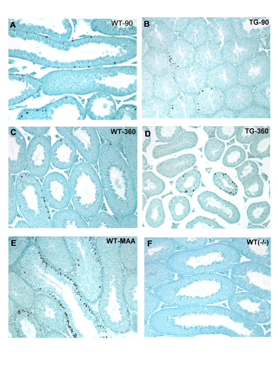
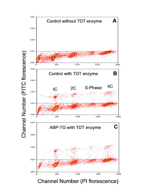
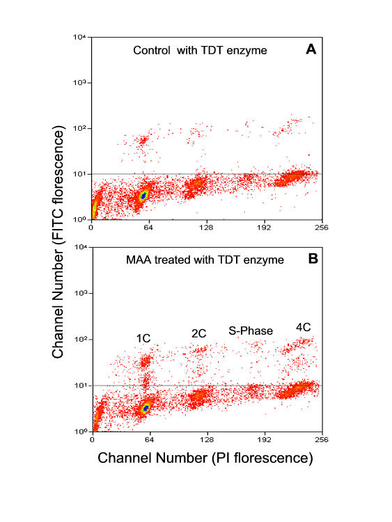
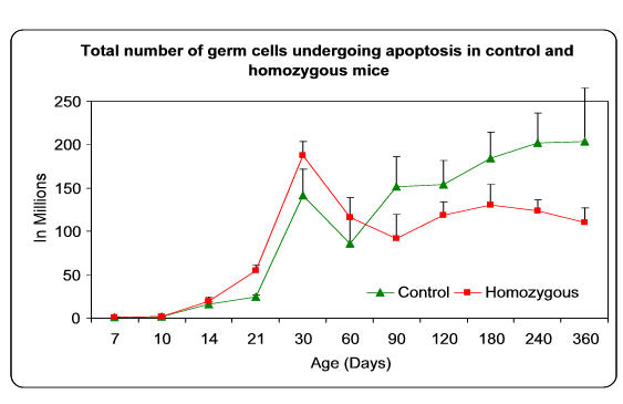
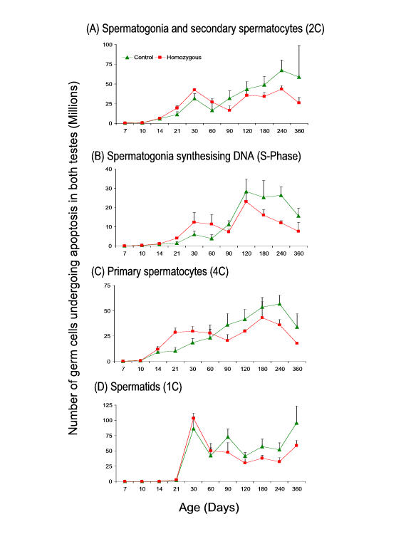
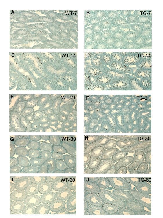
The number and type of testicular germ cells undergoing apoptosis in different age groups of mice (from 7 to 360 days of age) was determined and compared in age-matched wild type (WT) control and in a transgenic (TG) mice homozygous to rat androgen binding protein (ABP) using flow cytometry. Flow cytometric quantification revealed that the total number of germ cells undergoing apoptosis did not differ significantly in WT and TG mice up to Day 14. From Day 21 to Day 60, the number of germ cells undergoing apoptosis was consistently higher in TG than in WT mice. Starting from Day 90, the number of germ cells undergoing apoptosis in TG mice was lower than controls until Day 360. In 21-60 days old TG mice, spermatogonia, S-Phase cells, and primary spermatocytes are the cell types undergoing apoptosis at significantly greater numbers than those in WT mice. However, starting from day 60, the total number of spermatids undergoing apoptosis was significantly lower in TG mice than in age-matched WT controls. TdT-mediated dUTP-biotin nick end labeling (TUNEL) in testicular sections from TG mice of 21 and 30 days of age confirmed the presence of increased numbers of apoptotic germ cells compared to their age matched controls. These data indicate that the continuous presence of greater than physiological concentrations of ABP in the mouse testis has a biphasic effect on the frequency of apoptosis in germ cells. The initial pre-pubertal increase in testicular germ cell apoptosis may result from direct or indirect actions of ABP and is likely to determine the subsequent life-death balance of germ cell populations in TG mice, whereas the subsequent reduction may result from maturation depletion. A wave of apoptosis during the pre-pubertal period is required for normal spermatogenesis to develop, and our data indicate that this apoptotic wave may be regulated by ABP and/or androgens.
利用流式细胞术确定并比较了不同年龄组(7至360日龄)小鼠睾丸生殖细胞凋亡的数量和类型,这些小鼠包括年龄匹配的野生型(WT)对照小鼠以及同型合子的大鼠雄激素结合蛋白(ABP)转基因(TG)小鼠。流式细胞术定量分析显示,在14日龄之前,WT和TG小鼠中发生凋亡的生殖细胞总数没有显著差异。从21日龄到60日龄,TG小鼠中发生凋亡的生殖细胞数量始终高于WT小鼠。从90日龄开始,TG小鼠中发生凋亡的生殖细胞数量低于对照组,直至360日龄。在21至60日龄的TG小鼠中,精原细胞、S期细胞和初级精母细胞发生凋亡的细胞类型数量显著多于WT小鼠。然而,从60日龄开始,TG小鼠中发生凋亡的精子细胞总数显著低于年龄匹配的WT对照小鼠。对21日龄和30日龄TG小鼠睾丸切片进行的TdT介导的dUTP生物素缺口末端标记(TUNEL)证实,与年龄匹配的对照相比,凋亡生殖细胞数量增加。这些数据表明,小鼠睾丸中持续存在高于生理浓度的ABP对生殖细胞凋亡频率具有双相作用。青春期前睾丸生殖细胞凋亡的最初增加可能是ABP直接或间接作用的结果,并且可能决定了TG小鼠中生殖细胞群体随后的生死平衡,而随后的减少可能是由于成熟耗竭。青春期前的凋亡浪潮是正常精子发生发育所必需的,我们的数据表明,这一凋亡浪潮可能受ABP和/或雄激素调节。