• 文献检索
  • 文档翻译
  • 深度研究
  • 学术资讯
  • Suppr Zotero 插件Zotero 插件
  • 邀请有礼
  • 套餐&价格
  • 历史记录
应用&插件
Suppr Zotero 插件Zotero 插件浏览器插件Mac 客户端Windows 客户端微信小程序
定价
高级版会员购买积分包购买API积分包
服务
文献检索文档翻译深度研究API 文档MCP 服务
关于我们
关于 Suppr公司介绍联系我们用户协议隐私条款
关注我们

Suppr 超能文献

核心技术专利:CN118964589B侵权必究
粤ICP备2023148730 号-1Suppr @ 2026

文献检索

告别复杂PubMed语法,用中文像聊天一样搜索,搜遍4000万医学文献。AI智能推荐,让科研检索更轻松。

立即免费搜索

文件翻译

保留排版,准确专业,支持PDF/Word/PPT等文件格式,支持 12+语言互译。

免费翻译文档

深度研究

AI帮你快速写综述,25分钟生成高质量综述,智能提取关键信息,辅助科研写作。

立即免费体验

人糖尿病角膜在器官培养中保留了与正常角膜在伤口愈合、基底膜、整合素和基质金属蛋白酶-10方面的差异。

Human diabetic corneas preserve wound healing, basement membrane, integrin and MMP-10 differences from normal corneas in organ culture.

作者信息

Kabosova Andrea, Kramerov Andrei A, Aoki Annette M, Murphy Gillian, Zieske James D, Ljubimov Alexander V

机构信息

Ophthalmology Research Laboratories, Cedars-Sinai Medical Center, Burns and Allen Research Institute, Los Angeles, CA 90048, USA.

出版信息

Exp Eye Res. 2003 Aug;77(2):211-7. doi: 10.1016/s0014-4835(03)00111-8.

DOI:10.1016/s0014-4835(03)00111-8
PMID:12873452
原文链接:https://pmc.ncbi.nlm.nih.gov/articles/PMC2909880/
Abstract

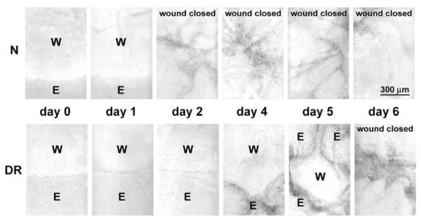
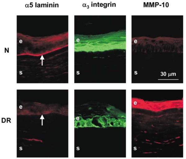
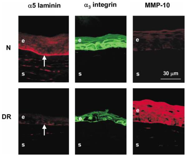
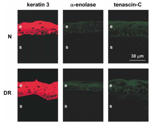
The authors have previously documented decreased epithelial basement membrane (BM) components and alpha3beta1 epithelial integrin, and increased expression of matrix metalloproteinase (MMP)-10 in corneas of patients with diabetic retinopathy (DR) compared to normal corneas. The purpose of this study was to examine if organ-cultured DR corneas exhibited the same alterations in wound healing and diabetic marker distribution as the autopsy DR corneas. Twenty normal and 17 DR corneas were organ-cultured in serum-free medium over agar-collagen gel at the air-liquid interface for up to 45 days. Circular 5 mm central epithelial wounds were made with n-heptanol, the procedure that will preserve fragile diabetic corneal BM. Wound healing was monitored microscopically every 12 hr. Distribution of diabetic corneal epithelial markers including laminin-10 alpha5 chain, nidogen-1/entactin, integrin alpha3beta1, and MMP-10, was examined by immunofluorescence. Normal corneas healed the central epithelial defect within 3 days (mean=2.3 days), whereas DR corneas on average healed about two times slower (mean=4.5 days). In wounded and completely healed organ-cultured corneas, the patterns of studied markers were the same as in the unwounded organ-cultured corneas. This concerned both normal and DR corneas. As in vivo, normal organ-cultured corneas had continuous staining for laminin-10 and nidogen-1/entactin in the epithelial BM, strong and homogeneous staining for both chains of alpha3beta1 integrin in epithelial cells, and little if any staining for MMP-10. Organ-cultured DR corneas also had marker patterns specific for in vivo DR corneas: interrupted to no staining for laminin-10 and nidogen-1/entactin in the epithelial BM, areas of weak or disorganized alpha3beta1 integrin in epithelial cells, and significant MMP-10 staining in the epithelium and keratocytes. Fibrotic extracellular matrix and myofibroblast markers were largely absent. Thus, epithelial wound healing was much slower in organ-cultured DR corneas than in normal corneas, in complete accordance with clinical data in diabetic patients. DR corneas in organ culture preserved the same marker abnormalities as in vivo. The marker distribution was unchanged in wounded and healed organ-cultured corneas, compared to unwounded corneas. The established corneal organ culture provides an adequate system for elucidating mechanisms of epithelial alterations in human DR corneas.

摘要

作者之前已证明,与正常角膜相比,糖尿病视网膜病变(DR)患者角膜的上皮基底膜(BM)成分和α3β1上皮整合素减少,基质金属蛋白酶(MMP)-10的表达增加。本研究的目的是检验器官培养的DR角膜在伤口愈合和糖尿病标志物分布方面是否表现出与尸检DR角膜相同的改变。将20只正常角膜和17只DR角膜在气液界面的琼脂-胶原凝胶上于无血清培养基中进行器官培养,长达45天。用正庚醇制作5mm圆形中央上皮伤口,该方法可保留脆弱的糖尿病角膜BM。每12小时通过显微镜监测伤口愈合情况。通过免疫荧光检查糖尿病角膜上皮标志物的分布,包括层粘连蛋白-10α5链、巢蛋白-1/内动蛋白、整合素α3β1和MMP-10。正常角膜在3天内(平均=2.3天)愈合中央上皮缺损,而DR角膜平均愈合速度慢约两倍(平均=4.5天)。在受伤和完全愈合的器官培养角膜中,所研究标志物的模式与未受伤的器官培养角膜相同。正常角膜和DR角膜均如此。与体内情况一样,正常器官培养角膜的上皮BM中层粘连蛋白-10和巢蛋白-1/内动蛋白呈连续染色,上皮细胞中α3β1整合素的两条链呈强而均匀的染色,MMP-10几乎无染色。器官培养的DR角膜也具有体内DR角膜特有的标志物模式:上皮BM中层粘连蛋白-10和巢蛋白-1/内动蛋白呈间断至无染色,上皮细胞中α3β1整合素呈弱或紊乱区域,上皮和角膜细胞中有明显的MMP-10染色。纤维化细胞外基质和成肌纤维细胞标志物基本不存在。因此,器官培养的DR角膜上皮伤口愈合比正常角膜慢得多,这与糖尿病患者的临床数据完全一致。器官培养中的DR角膜保留了与体内相同的标志物异常。与未受伤角膜相比,受伤和愈合的器官培养角膜中的标志物分布没有变化。已建立的角膜器官培养为阐明人类DR角膜上皮改变的机制提供了一个合适的系统。

https://cdn.ncbi.nlm.nih.gov/pmc/blobs/e8cc/2909880/fbdf52e99cfe/nihms-199811-f0004.jpg
https://cdn.ncbi.nlm.nih.gov/pmc/blobs/e8cc/2909880/27904fd3d948/nihms-199811-f0001.jpg
https://cdn.ncbi.nlm.nih.gov/pmc/blobs/e8cc/2909880/de52fea5d257/nihms-199811-f0002.jpg
https://cdn.ncbi.nlm.nih.gov/pmc/blobs/e8cc/2909880/2b504450e433/nihms-199811-f0003.jpg
https://cdn.ncbi.nlm.nih.gov/pmc/blobs/e8cc/2909880/fbdf52e99cfe/nihms-199811-f0004.jpg

相似文献

1
Human diabetic corneas preserve wound healing, basement membrane, integrin and MMP-10 differences from normal corneas in organ culture.人糖尿病角膜在器官培养中保留了与正常角膜在伤口愈合、基底膜、整合素和基质金属蛋白酶-10方面的差异。
Exp Eye Res. 2003 Aug;77(2):211-7. doi: 10.1016/s0014-4835(03)00111-8.
2
Human corneal epithelial basement membrane and integrin alterations in diabetes and diabetic retinopathy.糖尿病及糖尿病视网膜病变中人类角膜上皮基底膜和整合素的改变
J Histochem Cytochem. 1998 Sep;46(9):1033-41. doi: 10.1177/002215549804600907.
3
Overexpression of matrix metalloproteinase-10 and matrix metalloproteinase-3 in human diabetic corneas: a possible mechanism of basement membrane and integrin alterations.基质金属蛋白酶-10和基质金属蛋白酶-3在人糖尿病角膜中的过表达:基底膜和整合素改变的一种可能机制。
Am J Pathol. 2001 Feb;158(2):723-34. doi: 10.1016/S0002-9440(10)64015-1.
4
Enhanced wound healing, kinase and stem cell marker expression in diabetic organ-cultured human corneas upon MMP-10 and cathepsin F gene silencing.经 MMP-10 和组织蛋白酶 F 基因沉默后糖尿病器官培养人眼角膜的伤口愈合增强、激酶和干细胞标志物表达。
Invest Ophthalmol Vis Sci. 2013 Dec 17;54(13):8172-80. doi: 10.1167/iovs.13-13233.
5
Normalization of wound healing and stem cell marker patterns in organ-cultured human diabetic corneas by gene therapy of limbal cells.通过角膜缘细胞基因治疗使器官培养的人糖尿病角膜伤口愈合和干细胞标志物模式正常化。
Exp Eye Res. 2014 Dec;129:66-73. doi: 10.1016/j.exer.2014.10.022. Epub 2014 Oct 30.
6
Adenovirus-driven overexpression of proteinases in organ-cultured normal human corneas leads to diabetic-like changes.腺病毒驱动的在器官培养的正常人类角膜中过表达蛋白酶导致类似糖尿病的改变。
Brain Res Bull. 2010 Feb 15;81(2-3):262-72. doi: 10.1016/j.brainresbull.2009.10.007. Epub 2009 Oct 12.
7
Epithelial basement membrane proteins perlecan and nidogen-2 are up-regulated in stromal cells after epithelial injury in human corneas.人角膜上皮损伤后,基质细胞中的上皮基底膜蛋白核心蛋白聚糖和巢蛋白-2表达上调。
Exp Eye Res. 2015 May;134:33-8. doi: 10.1016/j.exer.2015.03.016. Epub 2015 Mar 19.
8
Proteinase and growth factor alterations revealed by gene microarray analysis of human diabetic corneas.通过基因芯片分析人糖尿病角膜揭示的蛋白酶和生长因子改变
Invest Ophthalmol Vis Sci. 2005 Oct;46(10):3604-15. doi: 10.1167/iovs.04-1507.
9
Normalization of wound healing and diabetic markers in organ cultured human diabetic corneas by adenoviral delivery of c-Met gene.腺病毒介导的 c-Met 基因转导对人糖尿病角膜器官培养中创面愈合和糖尿病标志物的正常化作用。
Invest Ophthalmol Vis Sci. 2010 Apr;51(4):1970-80. doi: 10.1167/iovs.09-4569. Epub 2009 Nov 20.
10
Abnormalities of the extracellular matrix in keratoconus corneas.圆锥角膜角膜细胞外基质的异常。
Cornea. 1997 May;16(3):345-51.

引用本文的文献

1
IFITM1/OVOL1 Axis Is a Novel Regulator of the Expansion of the Limbal Epithelial Stem/Early Transient Amplifying Cell Population.IFITM1/OVOL1轴是角膜缘上皮干细胞/早期短暂增殖细胞群扩增的新型调节因子。
FASEB J. 2025 May 31;39(10):e70648. doi: 10.1096/fj.202500783R.
2
Lack of Elevated Expression of TGFβ3 Contributes to the Delay of Epithelial Wound Healing in Diabetic Corneas.缺乏 TGFβ3 的高表达导致糖尿病角膜上皮伤口愈合延迟。
Invest Ophthalmol Vis Sci. 2024 Mar 5;65(3):35. doi: 10.1167/iovs.65.3.35.
3
Diabetic Keratopathy: Redox Signaling Pathways and Therapeutic Prospects.

本文引用的文献

1
Cell-matrix and cell-cell interactions during corneal epithelial wound healing.
Prog Retin Eye Res. 2003 Mar;22(2):113-33. doi: 10.1016/s1350-9462(02)00042-3.
2
Naltrexone, an opioid antagonist, facilitates reepithelialization of the cornea in diabetic rat.纳曲酮是一种阿片类拮抗剂,可促进糖尿病大鼠角膜的再上皮化。
Diabetes. 2002 Oct;51(10):3055-62. doi: 10.2337/diabetes.51.10.3055.
3
Extracellular matrix and wound healing.细胞外基质与伤口愈合
Curr Opin Ophthalmol. 2001 Aug;12(4):237-41. doi: 10.1097/00055735-200108000-00001.
糖尿病性角膜病变:氧化还原信号通路与治疗前景
Antioxidants (Basel). 2024 Jan 18;13(1):120. doi: 10.3390/antiox13010120.
4
Oxidative stress in the eye and its role in the pathophysiology of ocular diseases.眼睛中的氧化应激及其在眼部疾病病理生理学中的作用。
Redox Biol. 2023 Dec;68:102967. doi: 10.1016/j.redox.2023.102967. Epub 2023 Nov 18.
5
Limbal Epithelial Stem Cells in the Diabetic Cornea.糖尿病角膜中的角膜缘上皮干细胞。
Cells. 2023 Oct 16;12(20):2458. doi: 10.3390/cells12202458.
6
Chronic Hyperglycemia Compromises Mitochondrial Function in Corneal Epithelial Cells: Implications for the Diabetic Cornea.慢性高血糖损害角膜上皮细胞的线粒体功能:对糖尿病角膜的影响。
Cells. 2022 Aug 18;11(16):2567. doi: 10.3390/cells11162567.
7
Potential underlying genetic associations between keratoconus and diabetes mellitus.圆锥角膜与糖尿病之间潜在的潜在遗传关联。
Adv Ophthalmol Pract Res. 2021 Nov;1(1). doi: 10.1016/j.aopr.2021.100005. Epub 2021 Sep 4.
8
Cellular Contractility Profiles of Human Diabetic Corneal Stromal Cells.人糖尿病性角膜基质细胞的细胞收缩性特征。
Anal Cell Pathol (Amst). 2021 Jun 4;2021:9913210. doi: 10.1155/2021/9913210. eCollection 2021.
9
Systemic diseases and the cornea.系统性疾病与角膜。
Exp Eye Res. 2021 Mar;204:108455. doi: 10.1016/j.exer.2021.108455. Epub 2021 Jan 21.
10
Novel nanopolymer RNA therapeutics normalize human diabetic corneal wound healing and epithelial stem cells.新型纳米聚合物 RNA 疗法可使人类糖尿病性角膜伤口愈合和上皮干细胞正常化。
Nanomedicine. 2021 Feb;32:102332. doi: 10.1016/j.nano.2020.102332. Epub 2020 Nov 10.
4
Integrity of epithelium and endothelium in organ-cultured human corneas.器官培养的人角膜上皮和内皮的完整性。
Invest Ophthalmol Vis Sci. 2001 Jul;42(8):1757-61.
5
Overexpression of matrix metalloproteinase-10 and matrix metalloproteinase-3 in human diabetic corneas: a possible mechanism of basement membrane and integrin alterations.基质金属蛋白酶-10和基质金属蛋白酶-3在人糖尿病角膜中的过表达:基底膜和整合素改变的一种可能机制。
Am J Pathol. 2001 Feb;158(2):723-34. doi: 10.1016/S0002-9440(10)64015-1.
6
Matrix metalloproteinase activity is enhanced during corneal wound repair in high glucose condition.在高糖条件下,角膜伤口修复过程中基质金属蛋白酶活性增强。
Curr Eye Res. 2000 Aug;21(2):608-15.
7
Corneal organ culture model for assessing epithelial responses to surfactants.用于评估上皮细胞对表面活性剂反应的角膜器官培养模型
Toxicol Sci. 2000 Dec;58(2):306-14. doi: 10.1093/toxsci/58.2.306.
8
Response of capillary cell death to aminoguanidine predicts the development of retinopathy: comparison of diabetes and galactosemia.毛细血管细胞死亡对氨基胍的反应可预测视网膜病变的发展:糖尿病与半乳糖血症的比较。
Invest Ophthalmol Vis Sci. 2000 Nov;41(12):3972-8.
9
Glucose transporter 1 expression in corneal wound repair under high serum glucose level.高血糖水平下角膜伤口修复过程中葡萄糖转运蛋白1的表达
Jpn J Ophthalmol. 2000 Sep-Oct;44(5):470-4. doi: 10.1016/s0021-5155(00)00222-7.
10
Corneal structure and sensitivity in type 1 diabetes mellitus.1型糖尿病患者的角膜结构与敏感性
Invest Ophthalmol Vis Sci. 2000 Sep;41(10):2915-21.