• 文献检索
  • 文档翻译
  • 深度研究
  • 学术资讯
  • Suppr Zotero 插件Zotero 插件
  • 邀请有礼
  • 套餐&价格
  • 历史记录
应用&插件
Suppr Zotero 插件Zotero 插件浏览器插件Mac 客户端Windows 客户端微信小程序
定价
高级版会员购买积分包购买API积分包
服务
文献检索文档翻译深度研究API 文档MCP 服务
关于我们
关于 Suppr公司介绍联系我们用户协议隐私条款
关注我们

Suppr 超能文献

核心技术专利:CN118964589B侵权必究
粤ICP备2023148730 号-1Suppr @ 2026

文献检索

告别复杂PubMed语法,用中文像聊天一样搜索,搜遍4000万医学文献。AI智能推荐,让科研检索更轻松。

立即免费搜索

文件翻译

保留排版,准确专业,支持PDF/Word/PPT等文件格式,支持 12+语言互译。

免费翻译文档

深度研究

AI帮你快速写综述,25分钟生成高质量综述,智能提取关键信息,辅助科研写作。

立即免费体验

心电图检测左心房扩大。P波与经超声心动图测量的左心房大小的相关性。

Electrocardiographic detection of left atrial enlargement. Correlation of P wave with left atrial dimension by echocardiography.

作者信息

Chirife R, Feitosa G S, Frankl W S

出版信息

Br Heart J. 1975 Dec;37(12):1281-5. doi: 10.1136/hrt.37.12.1281.

DOI:10.1136/hrt.37.12.1281
PMID:131563
原文链接:https://pmc.ncbi.nlm.nih.gov/articles/PMC482954/
Abstract

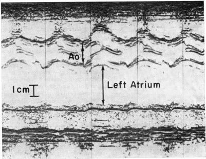
The validity of various electrocardiographic P wave measurements was tested in 48 patients by comparing them to left atrial dimensions determined by echocardiography (echo), a proved method of left atrial size estimation. Of all the measurements considered, only the width of the P wave (PW), the P terminal force in lead V1 (PV1), and the PW/PR segment ratio (PW/PR) showed statistically significant correlations with left atrial size measurements by echo, with r values of 0-746, 0-491, and 0-479, respectively. The results indicated that P widths in excess of 105 ms were present in all the patients who had left atria equal to or greater than 3-8 cm by echo and in 11 per cent of patients without atrial enlargement (false positives), and that when measurements were less than 105 ms left atrial enlargement was unlikely.

摘要

通过将各种心电图P波测量值与经超声心动图(echo)测定的左心房尺寸进行比较,对48例患者进行了各种心电图P波测量的有效性测试,超声心动图是一种已证实的左心房大小估计方法。在所有考虑的测量值中,只有P波宽度(PW)、V1导联的P波终末电势(PV1)以及PW/PR段比值(PW/PR)与超声心动图测量的左心房大小显示出统计学上的显著相关性,r值分别为0.746、0.491和0.479。结果表明,所有经超声心动图显示左心房等于或大于3.8 cm的患者以及11%无心房扩大的患者(假阳性)中均存在P波宽度超过105 ms的情况,而当测量值小于105 ms时,左心房扩大的可能性不大。

https://cdn.ncbi.nlm.nih.gov/pmc/blobs/1cbb/482954/d263c60cf918/brheartj00262-0079-a.jpg

相似文献

1
Electrocardiographic detection of left atrial enlargement. Correlation of P wave with left atrial dimension by echocardiography.心电图检测左心房扩大。P波与经超声心动图测量的左心房大小的相关性。
Br Heart J. 1975 Dec;37(12):1281-5. doi: 10.1136/hrt.37.12.1281.
2
Assessment of electrocardiographic criteria of left atrial enlargement.左心房扩大的心电图标准评估
Asian Cardiovasc Thorac Ann. 2018 May;26(4):273-276. doi: 10.1177/0218492318768131. Epub 2018 Mar 27.
3
Electrocardiographic left atrial enlargement--correlation with echo.心电图左心房增大——与超声心动图的相关性
J Assoc Physicians India. 1989 Aug;37(8):497-9.
4
Left atrial enlargement. Echocardiographic assessment of electrocardiographic criteria.
Circulation. 1976 Oct;54(4):553-7. doi: 10.1161/01.cir.54.4.553.
5
Sensitivity and specificity of commonly used electrocardiographic criteria for left atrial enlargement determined by M-mode echocardiography.通过M型超声心动图测定的常用心电图左心房扩大标准的敏感性和特异性。
Am J Cardiol. 1984 Mar 1;53(6):829-32. doi: 10.1016/0002-9149(84)90413-2.
6
The effect of left atrial histology and dimension on P wave morphology.左心房组织学和尺寸对P波形态的影响。
J Electrocardiol. 1983 Oct;16(4):363-6. doi: 10.1016/s0022-0736(83)80086-7.
7
Echocardiographic and electrocardiographic criteria for diagnosing left atrial enlargement.诊断左心房扩大的超声心动图和心电图标准。
South Med J. 1975 Feb;68(2):161-5. doi: 10.1097/00007611-197502000-00010.
8
[Diagnosis of left atrial enlargement with electrocardiogram. A misplaced reliance].
Cardiologia. 1994 Apr;39(4):247-52.
9
The non-invasive recognition of left atrial enlargement: comparison of electro- and echocardiographic measurements.左心房扩大的无创识别:心电图和超声心动图测量的比较。
Postgrad Med J. 1977 Jul;53(621):356-9. doi: 10.1136/pgmj.53.621.356.
10
Electrocardiographic diagnosis of left atrial enlargement in patients with mitral stenosis: the value of the P-wave area.
Acta Cardiol. 2003 Apr;58(2):139-41. doi: 10.2143/AC.58.2.2005266.

引用本文的文献

1
Correlating Left Atrial Enlargement and Left Ventricular Hypertrophy on ECG With Echocardiography and Cardiac Magnetic Resonance Imaging.心电图上左心房扩大和左心室肥厚与超声心动图及心脏磁共振成像的相关性
J Am Heart Assoc. 2025 Mar 18;14(6):e037772. doi: 10.1161/JAHA.124.037772. Epub 2025 Mar 13.
2
The Impact of Pulmonary Vein Anatomy on P-Wave Appearance during Sinus Rhythm: Cardiac Computed Tomography Study.肺静脉解剖结构对窦性心律时P波形态的影响:心脏计算机断层扫描研究
Diagnostics (Basel). 2023 Sep 11;13(18):2911. doi: 10.3390/diagnostics13182911.
3
The pivotal role of ECG in cardiomyopathies.

本文引用的文献

1
THE RELATIONSHIP OF LEFT ATRIAL PRESSURE AND VOLUME IN PATIENTS WITH HEART DISEASE.心脏病患者左心房压力与容积的关系
Am Heart J. 1964 May;67:635-42. doi: 10.1016/0002-8703(64)90334-5.
2
P-WAVE ANALYSIS IN VALVULAR HEART DISEASE.瓣膜性心脏病中的P波分析
Circulation. 1964 Feb;29:242-52. doi: 10.1161/01.cir.29.2.242.
3
A simple electrocardiographic indication of left atrial enlargement. A study of young patients with rheumatic heart disease.左心房扩大的一种简单心电图指征。对年轻风湿性心脏病患者的研究。
心电图在心肌病中的关键作用。
Front Cardiovasc Med. 2023 Jun 19;10:1178163. doi: 10.3389/fcvm.2023.1178163. eCollection 2023.
4
P wave area for quantitative electrocardiographic assessment of left atrial remodeling.用于左心房重构定量心电图评估的P波面积
PLoS One. 2014 Jun 5;9(6):e99178. doi: 10.1371/journal.pone.0099178. eCollection 2014.
5
P-wave dispersion: an indicator of cardiac autonomic dysfunction in children with neurocardiogenic syncope.P波离散度:神经心源性晕厥患儿心脏自主神经功能障碍的一个指标。
Pediatr Cardiol. 2014 Apr;35(4):596-600. doi: 10.1007/s00246-013-0825-y. Epub 2013 Oct 25.
6
P-wave morphology is associated with echocardiographic response to cardiac resynchronization therapy in MADIT-CRT patients.在MADIT-CRT患者中,P波形态与心脏再同步治疗的超声心动图反应相关。
Ann Noninvasive Electrocardiol. 2013 Nov;18(6):510-8. doi: 10.1111/anec.12121.
7
P-wave dispersion in children with acute rheumatic fever.急性风湿热患儿的P波离散度
Pediatr Cardiol. 2012 Jan;33(1):90-4. doi: 10.1007/s00246-011-0096-4. Epub 2011 Sep 6.
8
Utilization of Electrocardiographic P-wave Duration for AV Interval Optimization in Dual-Chamber Pacemakers.利用心电图P波时限优化双腔起搏器的房室间期
Indian Pacing Electrophysiol J. 2010 Sep 5;10(9):383-92.
9
The prognostic importance of isolated P-Wave abnormalities.孤立性 P 波异常的预后意义。
Clin Cardiol. 2010 Jun;33(6):E87-93. doi: 10.1002/clc.20628.
10
Usefulness of electrocardiographic parameters as compared with computed tomography measures of left atrial volume enlargement: from the ROMICAT trial.与计算机断层扫描测量的左心房容积增大相比,心电图参数的效用:来自ROMICAT试验
J Electrocardiol. 2011 Mar-Apr;44(2):257-64. doi: 10.1016/j.jelectrocard.2010.04.011. Epub 2010 May 27.
JAMA. 1963 Aug 3;185:358-62. doi: 10.1001/jama.1963.03060050036017.
4
Auricular overloadings; electrocardiographic analysis of 193 cases.
Am J Cardiol. 1959 Apr;3(4):453-71. doi: 10.1016/0002-9149(59)90366-2.
5
A method for the electrocardiographic recognition of atrial enlargement.一种用于心电图识别心房扩大的方法。
Circulation. 1958 May;17(5):882-9. doi: 10.1161/01.cir.17.5.882.
6
Relationship of the P wave to left atrial volume in rheumatic heart disease with mitral stenosis.风湿性心脏病二尖瓣狭窄时P波与左心房容积的关系
Am J Med Sci. 1958 Mar;235(3):290-6 passim. doi: 10.1097/00000441-195803000-00005.
7
A quantitative study of the electrocardiographic effects of atrial enlargement.心房扩大心电图效应的定量研究。
Am Heart J. 1957 Jan;53(1):55-64. doi: 10.1016/0002-8703(57)90383-6.
8
Estimation of left atrial size using ultrasound.
Am Heart J. 1969 Jul;78(1):43-52. doi: 10.1016/0002-8703(69)90257-9.
9
Electrocardiographic evidence of left atrial hypertension in acute myocardial infarction.急性心肌梗死时左心房高压的心电图证据
Circulation. 1973 Mar;47(3):493-8. doi: 10.1161/01.cir.47.3.493.
10
Prediction of left heart filling pressure and its sequential change in acute myocardial infarction from the terminal force of the P wave.根据P波终末力预测急性心肌梗死患者左心充盈压及其动态变化
Br Heart J. 1973 Feb;35(2):142-51. doi: 10.1136/hrt.35.2.142.