Lauredo Isabel T, Forteza Rosanna M, Botvinnikova Yelena, Abraham William M
Dept. of Research, Mount Sinai Medical Center, 4300 Alton Rd., Miami Beach, FL 33140, USA.
Am J Physiol Lung Cell Mol Physiol. 2004 Apr;286(4):L734-40. doi: 10.1152/ajplung.00129.2003. Epub 2003 Dec 5.
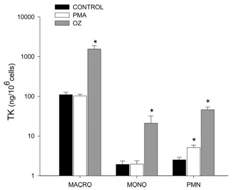
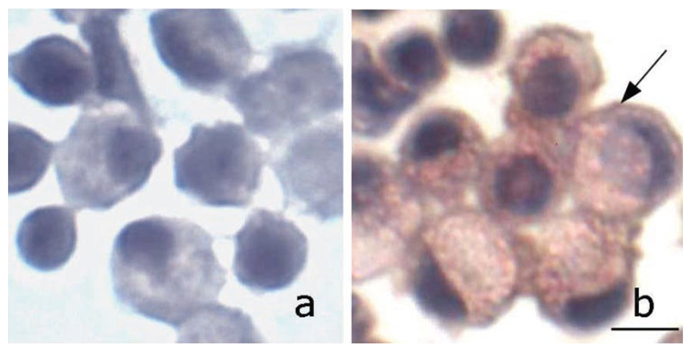
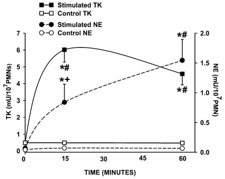
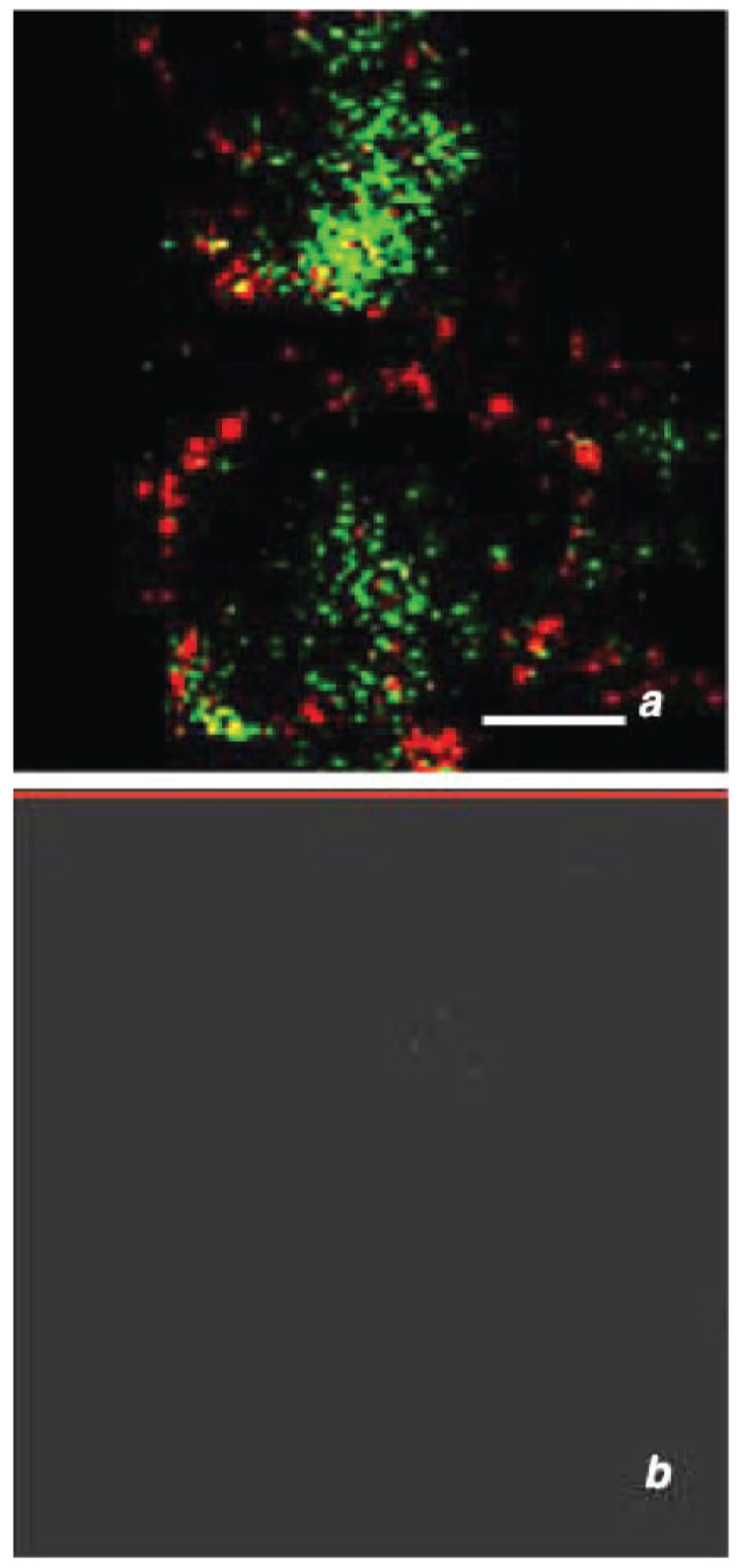
Lung tissue kallikrein (TK) is a serine proteinase that putatively plays a role in the pathophysiology of asthma by generating kallidin and bradykinin, mediators that contribute to airway hyperresponsiveness. In previous studies we observed biphasic increases in TK activity in bronchoalveolar lavage fluid following airway allergen challenge in allergic sheep. Although glandular TK is likely a major source of the initial increase in TK, the sources of the late increases in TK that are associated with the development of airway hyperresponsiveness may be dependent on activated resident and recruited inflammatory cells including alveolar macrophages (AMs) and neutrophils (PMNs). These cells increase concomitantly with the late increases in TK activity. To test this hypothesis, we obtained AMs from bronchoalveolar lavage fluid and PMNs and monocytes (precursors of AMs) from sheep blood and determined whether these cells contained TK and whether these same cells could release TK upon activation. Using confocal microscopy, immunocytochemical techniques, and enzyme activity assays, we found that all three cell types contained and secreted TK. All three cell types demonstrated basal release of TK, which could be increased after stimulation with zymosan. In addition, PMNs also released TK in the presence of phorbol ester, suggesting multiple secretory pathways in these cells. Furthermore, we showed that human monocytes also contain and secrete TK. We conclude that in the airways, monocytes, PMNs, and AMs may contribute to increased TK activity. Knowing the sources of TK in the airways could be important in understanding the mechanisms of inflammation that contribute to the pathophysiology of asthma and may help in the development of new therapies to control the disease.
肺组织激肽释放酶(TK)是一种丝氨酸蛋白酶,推测其通过生成缓激肽原和缓激肽在哮喘的病理生理学中发挥作用,这些介质会导致气道高反应性。在先前的研究中,我们观察到变应性绵羊气道过敏原激发后支气管肺泡灌洗液中TK活性呈双相增加。虽然腺性TK可能是TK最初增加的主要来源,但与气道高反应性发展相关的TK后期增加的来源可能取决于活化的驻留和募集的炎症细胞,包括肺泡巨噬细胞(AMs)和中性粒细胞(PMNs)。这些细胞与TK活性的后期增加同时增加。为了验证这一假设,我们从支气管肺泡灌洗液中获取AMs,从绵羊血液中获取PMNs和单核细胞(AMs的前体),并确定这些细胞是否含有TK,以及这些相同的细胞在活化后是否能释放TK。使用共聚焦显微镜、免疫细胞化学技术和酶活性测定,我们发现所有三种细胞类型都含有并分泌TK。所有三种细胞类型都表现出TK的基础释放,用酵母聚糖刺激后可增加。此外,PMNs在佛波酯存在的情况下也释放TK,表明这些细胞中有多种分泌途径。此外,我们还表明人类单核细胞也含有并分泌TK。我们得出结论,在气道中,单核细胞、PMNs和AMs可能导致TK活性增加。了解气道中TK的来源对于理解导致哮喘病理生理学的炎症机制可能很重要,并且可能有助于开发控制该疾病的新疗法。