Rochefort Gaël Y, Vaudin Pascal, Bonnet Nicolas, Pages Jean-Christophe, Domenech Jorge, Charbord Pierre, Eder Véronique
LABPART-EA3852, IFR135, Université François Rabelais, Faculté de Médecine, 10 Boulevard Tonnellé, 370032 TOURS, France.
Respir Res. 2005 Oct 27;6(1):125. doi: 10.1186/1465-9921-6-125.
Bone marrow (BM) cells are promising tools for vascular therapies. Here, we focused on the possibility of targeting the hypoxia-induced pulmonary artery hypertension remodeling with systemic delivery of BM-derived mesenchymal stem cells (MSCs) into non-irradiated rats.
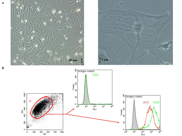
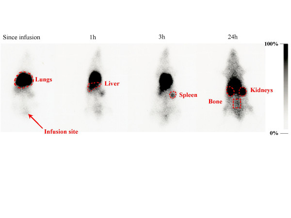
Six-week-old Wistar rats were exposed to 3-week chronic hypoxia leading to pulmonary artery wall remodeling. Domiciliation of adhesive BM-derived CD45- CD73+ CD90+ MSCs was first studied after a single intravenous infusion of Indium-111-labeled MSCs followed by whole body scintigraphies and autoradiographies of different harvested organs. In a second set of experiments, enhanced-GFP labeling allowed to observe distribution at later times using sequential infusions during the 3-week hypoxia exposure.
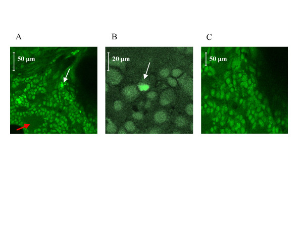
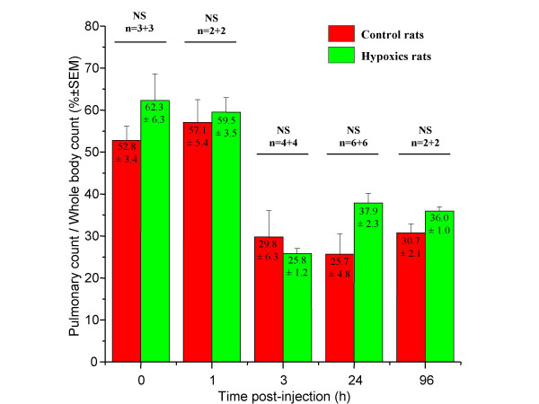
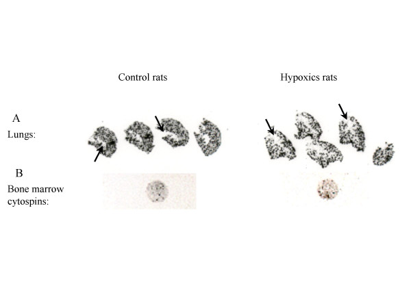
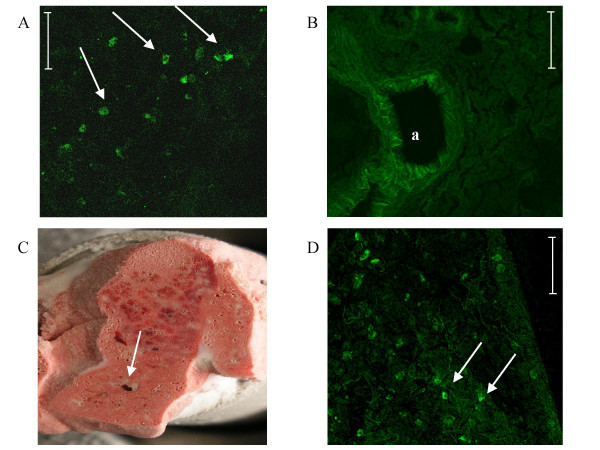
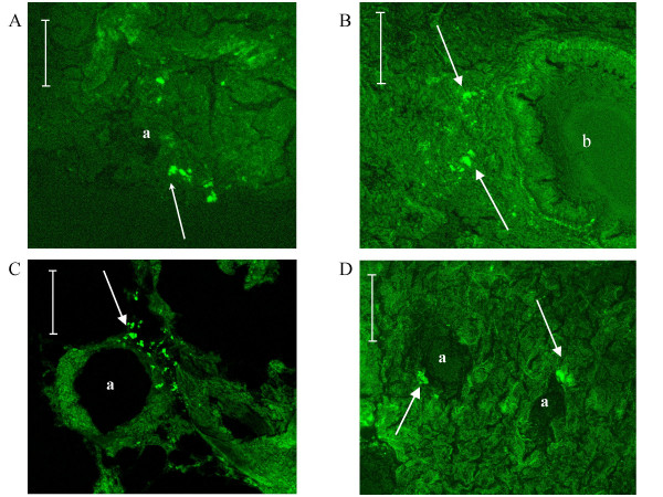
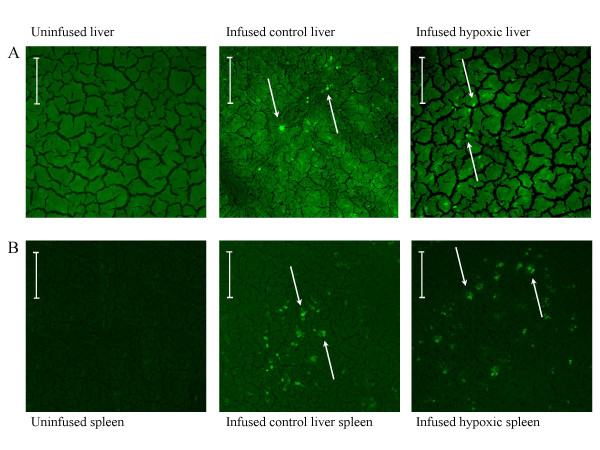
A 30% pulmonary retention was observed by scintigraphies and no differences were observed in the global repartition between hypoxic and control groups. Intrapulmonary radioactivity repartition was homogenous in both groups, as shown by autoradiographies. BM-derived GFP-labeled MSCs were observed with a global repartition in liver, in spleen, in lung parenchyma and rarely in the adventitial layer of remodeled vessels. Furthermore this global repartition was not modified by hypoxia. Interestingly, these cells displayed in vivo bone marrow homing, proving a preservation of their viability and function. Bone marrow homing of GFP-labeled MSCs was increased in the hypoxic group.
Adhesive BM-derived CD45- CD73+ CD90+ MSCs are not integrated in the pulmonary arteries remodeled media after repeated intravenous infusions in contrast to previously described in systemic vascular remodeling or with endothelial progenitor cells infusions.
骨髓细胞是血管治疗的有前景的工具。在此,我们聚焦于通过将骨髓来源的间充质干细胞(MSCs)全身递送至未受照射的大鼠体内,来靶向缺氧诱导的肺动脉高压重塑的可能性。
六周龄的Wistar大鼠暴露于3周的慢性缺氧环境,导致肺动脉壁重塑。在单次静脉输注铟-111标记的MSCs后,首先通过全身闪烁显像和不同收获器官的放射自显影研究黏附性骨髓来源的CD45-CD73+CD90+MSCs的归巢情况。在第二组实验中,增强型绿色荧光蛋白(GFP)标记允许在3周缺氧暴露期间通过连续输注在后期观察分布情况。
闪烁显像观察到肺部有30%的滞留,缺氧组和对照组之间的整体分布没有差异。放射自显影显示两组肺内放射性分布均均匀。观察到骨髓来源的GFP标记的MSCs在肝脏、脾脏、肺实质中整体分布,很少出现在重塑血管的外膜层。此外,这种整体分布不受缺氧影响。有趣的是,这些细胞在体内表现出骨髓归巢,证明其活力和功能得以保留。缺氧组中GFP标记的MSCs的骨髓归巢增加。
与先前在全身血管重塑或内皮祖细胞输注中所描述的情况不同,黏附性骨髓来源的CD45-CD73+CD90+MSCs在重复静脉输注后未整合到重塑的肺动脉中膜。