Eftekhari Mohammad, Assadi Majid, Kazemi Majid, Saghari Mohsen, Esfahani Armaghan Fard, Sichani Babak Fallahi, Gholamrezanezhad Ali, Beiki Davood
Research Institute for Nuclear Medicine, Tehran University of Medical Sciences, Shariati hospital, Northern Kargar St, 14114 Tehran, Iran.
BMC Nucl Med. 2005 Nov 28;5:6. doi: 10.1186/1471-2385-5-6.
Most olfactory testings are subjective and since they depend upon the patients' response, they are prone to false positive results. The aim of this study was to use quantitative brain perfusion SPECT in order to detect possible areas of brain activation in response to odorant stimulation in patients with post-traumatic impaired smell in comparison to a group of normal subjects.
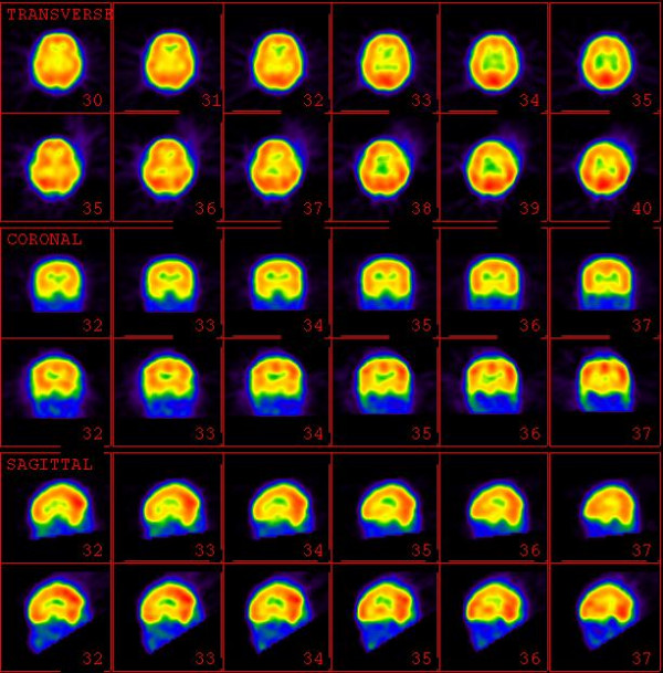
Fourteen patients with post-traumatic impaired smell and ten healthy controls were entered in this prospective study. All subjects underwent brain SPECT after intravenous injection of 740-MBq 99mTc-ECD and 48 hours later, the same procedure was repeated following olfactory stimulus (vanilla powder).
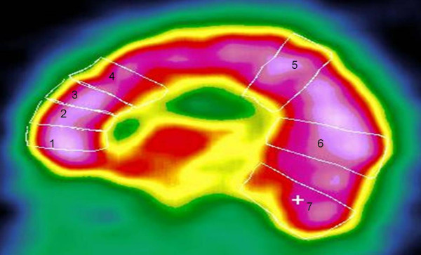
In most of seven regions of interest (Orbital Frontal Cortex, Inferior Frontal Pole, Superior Frontal Pole, Posterior Superior Frontal Lobe, Parasagittal Area, Occipital Pole, and Cerebellar area) the post-stimulation quantitative values show increased cortical perfusion being more pronounced in normal volunteers than the anosmic patients (except cerebellar areas and the right occipital pole). Maximal activation was observed in orbitofrontal regions (right+ 25.45% and left +25.47%).
Brain SPECT is a valuable imaging technique in the assessment of post-traumatic anosmia and could be competitive as an alternative to other imaging techniques, especially when functional MRI is unavailable or unsuitable. However, this procedure may benefit from complementary MRI or CT anatomical imaging.
大多数嗅觉测试是主观的,由于它们依赖于患者的反应,因此容易出现假阳性结果。本研究的目的是使用定量脑灌注单光子发射计算机断层扫描(SPECT),以检测创伤后嗅觉受损患者与一组正常受试者相比,在嗅觉刺激下可能出现的脑激活区域。
14名创伤后嗅觉受损患者和10名健康对照者纳入了这项前瞻性研究。所有受试者在静脉注射740MBq的99m锝-双半胱乙酯(99mTc-ECD)后接受脑SPECT检查,48小时后,在嗅觉刺激(香草粉)后重复相同的程序。
在大多数七个感兴趣区域(眶额皮质、额下极、额上极、额上叶后部、矢状旁区、枕极和小脑区域),刺激后的定量值显示皮质灌注增加,在正常志愿者中比嗅觉缺失患者更明显(小脑区域和右侧枕极除外)。在眶额区域观察到最大激活(右侧+25.45%,左侧+25.47%)。
脑SPECT是评估创伤后嗅觉缺失的一种有价值的成像技术,作为其他成像技术的替代方法可能具有竞争力,特别是当功能磁共振成像不可用或不合适时。然而,该程序可能受益于补充的磁共振成像或CT解剖成像。