Pentecost Mickey, Otto Glen, Theriot Julie A, Amieva Manuel R
Department of Microbiology and Immunology, Stanford University, Stanford, California, USA.
PLoS Pathog. 2006 Jan;2(1):e3. doi: 10.1371/journal.ppat.0020003. Epub 2006 Jan 27.
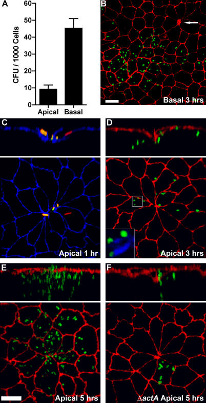
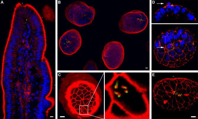
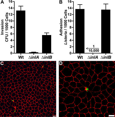
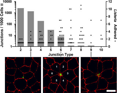
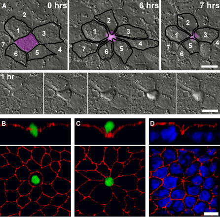
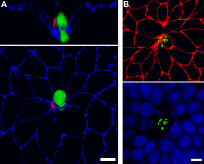
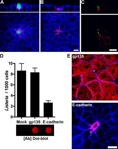
Listeria monocytogenes causes invasive disease by crossing the intestinal epithelial barrier. This process depends on the interaction between the bacterial surface protein Internalin A and the host protein E-cadherin, located below the epithelial tight junctions at the lateral cell-to-cell contacts. We used polarized MDCK cells as a model epithelium to determine how L. monocytogenes breaches the tight junctions to gain access to this basolateral receptor protein. We determined that L. monocytogenes does not actively disrupt the tight junctions, but finds E-cadherin at a morphologically distinct subset of intercellular junctions. We identified these sites as naturally occurring regions where single senescent cells are expelled and detached from the epithelium by extrusion. The surrounding cells reorganize to form a multicellular junction that maintains epithelial continuity. We found that E-cadherin is transiently exposed to the lumenal surface at multicellular junctions during and after cell extrusion, and that L. monocytogenes takes advantage of junctional remodeling to adhere to and subsequently invade the epithelium. In intact epithelial monolayers, an anti-E-cadherin antibody specifically decorates multicellular junctions and blocks L. monocytogenes adhesion. Furthermore, an L. monocytogenes mutant in the Internalin A gene is completely deficient in attachment to the epithelial apical surface and is unable to invade. We hypothesized that L. monocytogenes utilizes analogous extrusion sites for epithelial invasion in vivo. By infecting rabbit ileal loops, we found that the junctions at the cell extrusion zone of villus tips are the specific target for L. monocytogenes adhesion and invasion. Thus, L. monocytogenes exploits the dynamic nature of epithelial renewal and junctional remodeling to breach the intestinal barrier.
单核细胞增生李斯特菌通过穿越肠道上皮屏障引发侵袭性疾病。这一过程依赖于细菌表面蛋白内化素A与宿主蛋白E-钙黏蛋白之间的相互作用,E-钙黏蛋白位于上皮细胞紧密连接下方的细胞间侧向连接处。我们使用极化的MDCK细胞作为模型上皮细胞,以确定单核细胞增生李斯特菌如何突破紧密连接从而接触到这种基底外侧受体蛋白。我们确定单核细胞增生李斯特菌不会主动破坏紧密连接,而是在细胞间连接的一个形态学上独特的亚群中找到E-钙黏蛋白。我们将这些位点确定为自然发生的区域,在此单个衰老细胞通过挤压从上皮细胞中排出并脱离。周围的细胞重新组织形成一个维持上皮连续性的多细胞连接。我们发现,在细胞挤压期间及之后,E-钙黏蛋白会在多细胞连接处短暂暴露于管腔表面,并且单核细胞增生李斯特菌利用连接重塑来黏附并随后侵入上皮细胞。在完整的上皮单层中,抗E-钙黏蛋白抗体特异性地标记多细胞连接并阻断单核细胞增生李斯特菌的黏附。此外,内化素A基因的单核细胞增生李斯特菌突变体在上皮细胞顶端表面的附着完全缺陷,并且无法侵入。我们推测单核细胞增生李斯特菌在体内利用类似的挤压位点进行上皮细胞侵袭。通过感染兔回肠袢,我们发现绒毛尖端细胞挤压区的连接处是单核细胞增生李斯特菌黏附和侵袭的特定靶点。因此,单核细胞增生李斯特菌利用上皮更新和连接重塑的动态特性来突破肠道屏障。