Penido Norma de Oliveira, Borin Andrei, Fukuda Yotaka, Lion Cristina Navarro Santos
Federal University of Sao Paulo (UNIFESP), Escola Paulista de Medicina.
Braz J Otorhinolaryngol. 2005 Jul-Aug;71(4):410-4. doi: 10.1016/s1808-8694(15)31191-5. Epub 2005 Dec 15.
The knowledge of the relations between the noble and vital structures of temporal bone is still a great challenge for the otologic surgeon. The microscopic anatomic studies of the temporal bone are one of the greatest help to prevent lesions during surgical intervention.
To study the anatomic correlations between the carotid canal and the cochlea, and the occurrence of dehiscence of the carotid canal in the middle ear tympanic cavity.
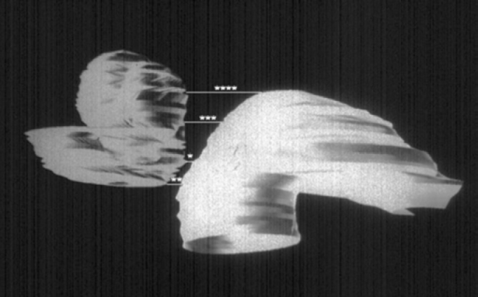
Microscopic study of 122 human temporal bones.
The average distance between the carotid canal and the cochlea were: the shortest distance, 1.05 mm; basal turn, 2.04 mm; middle turn, 2.32 mm; and apical turn, 5.70 mm. The occurrence of dehiscence of the carotid canal inside the tympanic cavity was 35.2%.
The small distances between the cochlea and carotid canal, and the high incidence of dehiscence in the tympanic cavity remind us that anatomical knowledge of the temporal bone is required for the best qualification of otologists.
颞骨的重要结构与生命结构之间的关系知识,对耳科外科医生来说仍是巨大挑战。颞骨的微观解剖学研究对防止手术干预期间的损伤有极大帮助。
研究颈动脉管与耳蜗之间的解剖学关联,以及中耳鼓室内颈动脉管裂缺的发生率。
对122块人类颞骨进行微观研究。
颈动脉管与耳蜗之间的平均距离为:最短距离1.05毫米;底转2.04毫米;中转2.32毫米;顶转5.70毫米。鼓室内颈动脉管裂缺的发生率为35.2%。
耳蜗与颈动脉管之间距离小,且鼓室内裂缺发生率高,这提醒我们,耳科医生要具备最佳资质,需要掌握颞骨的解剖学知识。