• 文献检索
  • 文档翻译
  • 深度研究
  • 学术资讯
  • Suppr Zotero 插件Zotero 插件
  • 邀请有礼
  • 套餐&价格
  • 历史记录
应用&插件
Suppr Zotero 插件Zotero 插件浏览器插件Mac 客户端Windows 客户端微信小程序
定价
高级版会员购买积分包购买API积分包
服务
文献检索文档翻译深度研究API 文档MCP 服务
关于我们
关于 Suppr公司介绍联系我们用户协议隐私条款
关注我们

Suppr 超能文献

核心技术专利:CN118964589B侵权必究
粤ICP备2023148730 号-1Suppr @ 2026

文献检索

告别复杂PubMed语法,用中文像聊天一样搜索,搜遍4000万医学文献。AI智能推荐,让科研检索更轻松。

立即免费搜索

文件翻译

保留排版,准确专业,支持PDF/Word/PPT等文件格式,支持 12+语言互译。

免费翻译文档

深度研究

AI帮你快速写综述,25分钟生成高质量综述,智能提取关键信息,辅助科研写作。

立即免费体验

相似文献

1
Expression of the gamma-aminobutyric acid (GABA) plasma membrane transporter-1 in monkey and human retina.γ-氨基丁酸(GABA)质膜转运体-1在猴和人视网膜中的表达。
Invest Ophthalmol Vis Sci. 2006 Apr;47(4):1682-90. doi: 10.1167/iovs.05-1117.
2
Multiple gamma-Aminobutyric acid plasma membrane transporters (GAT-1, GAT-2, GAT-3) in the rat retina.大鼠视网膜中的多种γ-氨基丁酸质膜转运体(GAT-1、GAT-2、GAT-3)
J Comp Neurol. 1996 Nov 11;375(2):212-24. doi: 10.1002/(SICI)1096-9861(19961111)375:2<212::AID-CNE3>3.0.CO;2-5.
3
Immunocytochemical localization of gamma-aminobutyric acid plasma membrane transporters in the tiger salamander retina.γ-氨基丁酸质膜转运体在虎螈视网膜中的免疫细胞化学定位。
J Comp Neurol. 1997 Dec 8;389(1):117-26. doi: 10.1002/(sici)1096-9861(19971208)389:1<117::aid-cne8>3.0.co;2-5.
4
Plasmalemmal and vesicular gamma-aminobutyric acid transporter expression in the developing mouse retina.发育中小鼠视网膜中质膜和囊泡γ-氨基丁酸转运体的表达
J Comp Neurol. 2009 Jan 1;512(1):6-26. doi: 10.1002/cne.21846.
5
Regulation of GABA content by glucose in the chick retina.鸡视网膜中葡萄糖对 GABA 含量的调节。
Exp Eye Res. 2013 Oct;115:206-15. doi: 10.1016/j.exer.2013.07.026. Epub 2013 Aug 3.
6
Expression of GAT-1, a high-affinity gamma-aminobutyric acid plasma membrane transporter in the rat retina.GAT-1(一种高亲和力γ-氨基丁酸质膜转运体)在大鼠视网膜中的表达。
J Comp Neurol. 1994 Jul 22;345(4):602-11. doi: 10.1002/cne.903450410.
7
Vesicular gamma-aminobutyric acid transporter expression in amacrine and horizontal cells.无长突细胞和水平细胞中囊泡型γ-氨基丁酸转运体的表达
J Comp Neurol. 2002 Apr 8;445(3):227-37. doi: 10.1002/cne.10166.
8
Cyan fluorescent protein expression in ganglion and amacrine cells in a thy1-CFP transgenic mouse retina.在thy1-CFP转基因小鼠视网膜的神经节细胞和无长突细胞中青色荧光蛋白的表达。
Mol Vis. 2008 Aug 25;14:1559-74.
9
Immunohistochemical localization of GABAA receptors in the retina of the new world primate Saimiri sciureus.γ-氨基丁酸A型受体在新大陆灵长类动物松鼠猴视网膜中的免疫组织化学定位。
Vis Neurosci. 1989;2(6):565-81. doi: 10.1017/s0952523800003503.
10
Distribution and synaptic connectivity of neuropeptide Y-immunoreactive amacrine cells in the rat retina.大鼠视网膜中神经肽Y免疫反应性无长突细胞的分布及突触连接
J Comp Neurol. 2002 May 6;446(3):219-34. doi: 10.1002/cne.10184.

引用本文的文献

1
Retinal VIP-amacrine cells: their development, structure, and function.视网膜 VIP 无长突细胞:它们的发育、结构和功能。
Eye (Lond). 2024 Apr;38(6):1065-1076. doi: 10.1038/s41433-023-02844-x. Epub 2023 Dec 8.
2
Starvation to Glucose Reprograms Development of Neurovascular Unit in Embryonic Retinal Cells.饥饿至葡萄糖重编程胚胎视网膜细胞中神经血管单元的发育。
Front Cell Dev Biol. 2021 Nov 18;9:726852. doi: 10.3389/fcell.2021.726852. eCollection 2021.
3
Vesicular Release of GABA by Mammalian Horizontal Cells Mediates Inhibitory Output to Photoreceptors.哺乳动物水平细胞以囊泡形式释放γ-氨基丁酸介导对光感受器的抑制性输出。
Front Cell Neurosci. 2020 Dec 1;14:600777. doi: 10.3389/fncel.2020.600777. eCollection 2020.
4
Differentiation of human ESCs to retinal ganglion cells using a CRISPR engineered reporter cell line.利用CRISPR工程改造的报告细胞系将人胚胎干细胞分化为视网膜神经节细胞。
Sci Rep. 2015 Nov 13;5:16595. doi: 10.1038/srep16595.
5
Histamine receptors of cones and horizontal cells in Old World monkey retinas.旧世界猴视网膜中的视锥细胞和水平细胞的组胺受体。
J Comp Neurol. 2012 Feb 15;520(3):528-43. doi: 10.1002/cne.22731.
6
Immunocytochemical evidence for SNARE protein-dependent transmitter release from guinea pig horizontal cells.免疫细胞化学证据表明 SNARE 蛋白依赖的豚鼠水平细胞递质释放。
Eur J Neurosci. 2010 Apr;31(8):1388-401. doi: 10.1111/j.1460-9568.2010.07181.x. Epub 2010 Apr 6.
7
Guinea pig horizontal cells express GABA, the GABA-synthesizing enzyme GAD 65, and the GABA vesicular transporter.豚鼠水平细胞表达 GABA、GABA 合成酶 GAD65 和 GABA 囊泡转运蛋白。
J Comp Neurol. 2010 May 15;518(10):1647-69. doi: 10.1002/cne.22294.
8
Twenty-one-base-pair insertion polymorphism creates an enhancer element and potentiates SLC6A1 GABA transporter promoter activity.21个碱基对的插入多态性产生一个增强子元件并增强SLC6A1γ-氨基丁酸转运体启动子活性。
Pharmacogenet Genomics. 2009 Jan;19(1):53-65. doi: 10.1097/FPC.0b013e328318b21a.
9
Plasmalemmal and vesicular gamma-aminobutyric acid transporter expression in the developing mouse retina.发育中小鼠视网膜中质膜和囊泡γ-氨基丁酸转运体的表达
J Comp Neurol. 2009 Jan 1;512(1):6-26. doi: 10.1002/cne.21846.
10
Cyan fluorescent protein expression in ganglion and amacrine cells in a thy1-CFP transgenic mouse retina.在thy1-CFP转基因小鼠视网膜的神经节细胞和无长突细胞中青色荧光蛋白的表达。
Mol Vis. 2008 Aug 25;14:1559-74.

本文引用的文献

1
Differential expression of the GABA transporters GAT-1 and GAT-3 in brains of rats, cats, monkeys and humans.大鼠、猫、猴及人类大脑中γ-氨基丁酸转运体GAT-1和GAT-3的差异表达
Cell Tissue Res. 2005 Jun;320(3):379-92. doi: 10.1007/s00441-004-0928-0. Epub 2005 Apr 9.
2
Nitric oxide stimulates gamma-aminobutyric acid release and inhibits glycine release in retina.一氧化氮刺激视网膜中γ-氨基丁酸的释放并抑制甘氨酸的释放。
J Comp Neurol. 2005 Mar 14;483(3):278-91. doi: 10.1002/cne.20416.
3
Blockade of the membranal GABA transporter potentiates GABAergic responses evoked in pyramidal cells by mossy fiber activation after seizures.癫痫发作后,阻断膜性γ-氨基丁酸(GABA)转运体可增强苔藓纤维激活在锥体细胞中诱发的GABA能反应。
Hippocampus. 2005;15(3):281-4. doi: 10.1002/hipo.20063.
4
GABA transporters in the mammalian cerebral cortex: localization, development and pathological implications.哺乳动物大脑皮层中的γ-氨基丁酸转运体:定位、发育及病理意义
Brain Res Brain Res Rev. 2004 Jul;45(3):196-212. doi: 10.1016/j.brainresrev.2004.03.003.
5
Perisynaptic localization of delta subunit-containing GABA(A) receptors and their activation by GABA spillover in the mouse dentate gyrus.含δ亚基的GABA(A)受体在小鼠齿状回突触周围的定位及其通过GABA外溢的激活。
J Neurosci. 2003 Nov 19;23(33):10650-61. doi: 10.1523/JNEUROSCI.23-33-10650.2003.
6
Transport-mediated synapses in the retina.视网膜中由转运介导的突触。
Physiol Rev. 2002 Oct;82(4):875-91. doi: 10.1152/physrev.00010.2002.
7
GAT-1 and reversible GABA transport in Bergmann glia in slices.伯格曼胶质细胞切片中的GAT-1与可逆性γ-氨基丁酸转运
J Neurophysiol. 2002 Sep;88(3):1407-19. doi: 10.1152/jn.2002.88.3.1407.
8
High-affinity GABA uptake in retinal glial (Müller) cells of the guinea pig: electrophysiological characterization, immunohistochemical localization, and modeling of efficiency.豚鼠视网膜神经胶质(穆勒)细胞中的高亲和力γ-氨基丁酸摄取:电生理特性、免疫组织化学定位及效率建模
Glia. 2002 Sep;39(3):217-28. doi: 10.1002/glia.10097.
9
GABA transporters regulate inhibition in the retina by limiting GABA(C) receptor activation.γ-氨基丁酸转运体通过限制γ-氨基丁酸C型受体的激活来调节视网膜中的抑制作用。
J Neurosci. 2002 Apr 15;22(8):3285-92. doi: 10.1523/JNEUROSCI.22-08-03285.2002.
10
Efficacy of background GABA uptake in rat hippocampal slices.大鼠海马切片中背景γ-氨基丁酸摄取的功效
Neuroreport. 2001 Jun 13;12(8):1593-6. doi: 10.1097/00001756-200106130-00016.

γ-氨基丁酸(GABA)质膜转运体-1在猴和人视网膜中的表达。

Expression of the gamma-aminobutyric acid (GABA) plasma membrane transporter-1 in monkey and human retina.

作者信息

Casini Giovanni, Rickman Dennis W, Brecha Nicholas C

机构信息

Dipartimento di Scienze Ambientali, Università della Tuscia, Viterbo, Italy.

出版信息

Invest Ophthalmol Vis Sci. 2006 Apr;47(4):1682-90. doi: 10.1167/iovs.05-1117.

DOI:10.1167/iovs.05-1117
PMID:16565409
原文链接:https://pmc.ncbi.nlm.nih.gov/articles/PMC3696021/
Abstract

PURPOSE

To determine the expression pattern of the predominant gamma-aminobutyric acid (GABA) plasma membrane transporter GAT-1 in Old World monkey (Macaca mulatta) and human retina.

METHODS

GAT-1 was localized in retinal sections by using immunohistochemical techniques with fluorescence and confocal microscopy. Double-labeling studies were performed with the GAT-1 antibody using antibodies to GABA, vasoactive intestinal polypeptide (VIP), tyrosine hydroxylase (TH), and the bipolar cell marker Mab115A10.

RESULTS

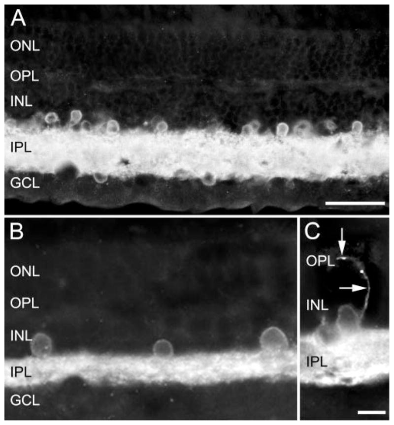
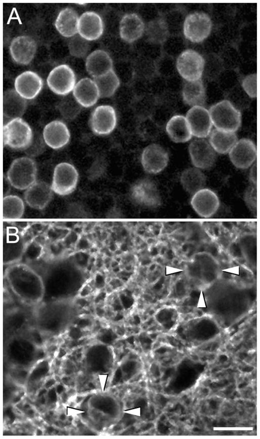
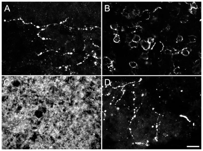
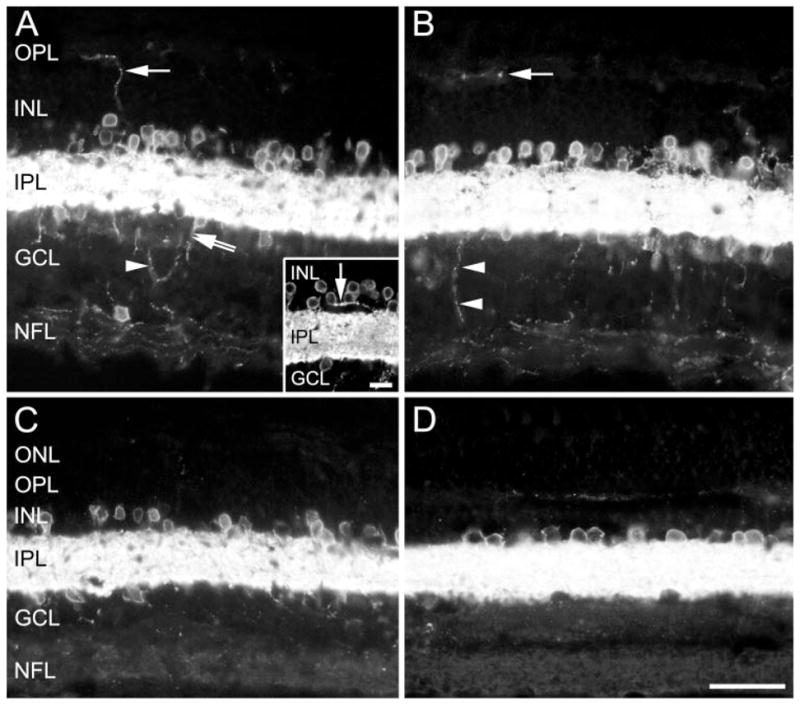
The pattern of GAT-1 immunostaining was similar in human and monkey retinas. Numerous small immunoreactive somata were in the inner nuclear layer (INL) and were present rarely in the inner plexiform layer (IPL) of all retinal regions. Medium GAT-1 somata were in the ganglion cell layer in the parafoveal and peripheral retinal regions. GAT-1 fibers were densely distributed throughout the IPL. Varicose processes, originating from both the IPL and somata in the INL, arborized in the outer plexiform layer (OPL), forming a sparse network in all retinal regions, except the fovea. Sparsely occurring GAT-1 processes were in the nerve fiber layer in parafoveal regions and near the optic nerve head but not in the optic nerve. In the INL, 99% of the GAT-1 somata contained GABA, and 66% of the GABA immunoreactive somata expressed GAT-1. GAT-1 immunoreactivity was in all VIP-containing cells, but it was absent in TH-immunoreactive amacrine cells and in Mab115A10 immunoreactive bipolar cells.

CONCLUSIONS

GAT-1 in primate retinas is expressed by amacrine and displaced amacrine cells. The predominant expression of GAT-1 in the inner retina is consistent with the idea that GABA transporters influence neurotransmission and thus participate in visual information processing in the retina.

摘要

目的

确定旧世界猴(猕猴)和人类视网膜中主要的γ-氨基丁酸(GABA)质膜转运体GAT-1的表达模式。

方法

采用免疫组织化学技术结合荧光和共聚焦显微镜,将GAT-1定位在视网膜切片中。使用针对GABA、血管活性肠肽(VIP)、酪氨酸羟化酶(TH)和双极细胞标志物Mab115A10的抗体,对GAT-1抗体进行双标记研究。

结果

人类和猴视网膜中GAT-1免疫染色模式相似。在内核层(INL)中有许多小的免疫反应性胞体,在所有视网膜区域的内网状层(IPL)中很少出现。中等大小的GAT-1胞体位于中央凹旁和周边视网膜区域的神经节细胞层。GAT-1纤维密集分布于整个IPL。起源于IPL和INL中胞体的曲张突起在外网状层(OPL)中分支,在除中央凹外的所有视网膜区域形成稀疏网络。在中央凹旁区域的神经纤维层和视神经乳头附近有少量GAT-1突起,但在视神经中没有。在INL中,99%的GAT-1胞体含有GABA,66%的GABA免疫反应性胞体表达GAT-1。GAT-1免疫反应性存在于所有含VIP的细胞中,但在TH免疫反应性无长突细胞和Mab115A10免疫反应性双极细胞中不存在。

结论

灵长类视网膜中的GAT-1由无长突细胞和移位无长突细胞表达。GAT-1在内视网膜中的主要表达与GABA转运体影响神经传递并因此参与视网膜视觉信息处理的观点一致。

https://cdn.ncbi.nlm.nih.gov/pmc/blobs/bf85/3696021/664600410fc5/nihms194736f8.jpg
https://cdn.ncbi.nlm.nih.gov/pmc/blobs/bf85/3696021/5d61d3047f4a/nihms194736f1.jpg
https://cdn.ncbi.nlm.nih.gov/pmc/blobs/bf85/3696021/74d277bb5697/nihms194736f2.jpg
https://cdn.ncbi.nlm.nih.gov/pmc/blobs/bf85/3696021/bd4b73943351/nihms194736f3.jpg
https://cdn.ncbi.nlm.nih.gov/pmc/blobs/bf85/3696021/00ffcc2ba0e5/nihms194736f4.jpg
https://cdn.ncbi.nlm.nih.gov/pmc/blobs/bf85/3696021/91c62493c415/nihms194736f5.jpg
https://cdn.ncbi.nlm.nih.gov/pmc/blobs/bf85/3696021/b280768099cb/nihms194736f6.jpg
https://cdn.ncbi.nlm.nih.gov/pmc/blobs/bf85/3696021/667a10205960/nihms194736f7.jpg
https://cdn.ncbi.nlm.nih.gov/pmc/blobs/bf85/3696021/664600410fc5/nihms194736f8.jpg