Voisard Rainer, Alan Mustafa, von Müller Lutz, Baur Regine, Hombach Vinzenz
Department of Internal Medicine II-Cardiology, University of Ulm, Robert-Kochstrasse 8, D-89081 Ulm, Germany.
BMC Cardiovasc Disord. 2006 Apr 4;6:14. doi: 10.1186/1471-2261-6-14.
The significant reduction of angiographic restenosis rates in the ISAR-SWEET study (intracoronary stenting and antithrombotic regimen: is abciximab a superior way to eliminate elevated thrombotic risk in diabetes) raises the question of whether abciximab acts on clopidogrel-independent mechanisms in suppressing neointimal hyperplasia. The current study investigates the direct effect of abciximab on ICAM-1 expression, migration and proliferation.
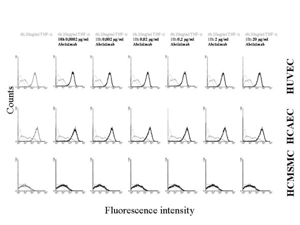
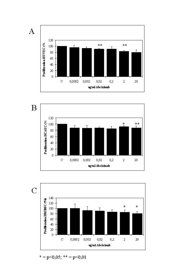
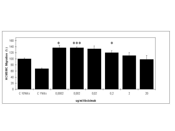
ICAM-1: Part I of the study investigates in cytoflow studies the effect of abciximab (0.0002, 0.002, 0.02, 0.2, 2.0, and 20.0 microg/ml) on TNF-alpha induced expression of intercellular adhesion molecule 1 (ICAM-1). Migration: Part II of the study explored the effect of abciximab (0.0002, 0.002, 0.02, 0.2, 2.0, and 20.0 microg/ml) on migration of HCMSMC over a period of 24 h. Proliferation: Part III of the study investigated the effect of abciximab (0.0002, 0.002, 0.02, 0.2, 2.0, and 20.0 microg/ml) on proliferation of HUVEC, HCAEC, and HCMSMC after an incubation period of 5 days.
ICAM-1: In human venous endothelial cells (HUVEC), human coronary endothelial cells (HCAEC) and human coronary medial smooth muscle cells (HCMSMC) no inhibitory or stimulatory effect on expression of ICAM-1 was detected. Migration: After incubation of HCMSMC with abciximab in concentrations of 0.0002-2 microg/ml a stimulatory effect on cell migration was detected, statistical significance was achieved after incubation with 0.002 microg/ml (p < 0.05), 0.002 microg/ml (p < 0.001), and 0.2 microg/ml (p < 0.05). Proliferation: Small but statistically significant antiproliferative effects of abciximab were detected after incubation of HUVEC (0.02 and 2.0 microg/ml; p = 0.01 and p < 0.01), HCAEC (2.0 and 20.0 microg/ml; p < 0.05 and p < 0,01), and HCMSMC (2.0 and 20.0 microg/ml; p < 0.05 and p < 0.05). The significant inhibition (SI) of cell proliferation found in HCAEC and HCMSMC was achieved with drug concentrations more than 10 times beyond the maximal plasma level (MPL), resulting in a SI/MPL-ratio > 1.
Thus, the anti-restenotic effects of systemically administered abciximab reported in the ISAR-SWEET-study were not caused by a direct inhibitory effect on ICAM-1 expression, migration or proliferation.
ISAR-SWEET研究(冠状动脉内支架置入术和抗血栓治疗方案:阿昔单抗是否是消除糖尿病患者血栓形成风险升高的更佳方法)中血管造影再狭窄率显著降低,这引发了一个问题,即阿昔单抗在抑制内膜增生方面是否通过不依赖氯吡格雷的机制发挥作用。本研究调查了阿昔单抗对细胞间黏附分子-1(ICAM-1)表达、迁移和增殖的直接影响。
ICAM-1:研究的第一部分在细胞流式研究中调查了阿昔单抗(0.0002、0.002、0.02、0.2、2.0和20.0微克/毫升)对肿瘤坏死因子-α诱导的细胞间黏附分子1(ICAM-1)表达的影响。迁移:研究的第二部分探讨了阿昔单抗(0.0002、0.002、0.02、0.2、2.0和20.0微克/毫升)在24小时内对人冠状动脉平滑肌细胞(HCMSMC)迁移的影响。增殖:研究的第三部分调查了阿昔单抗(0.0002、0.002、0.02、0.2、2.0和20.0微克/毫升)在孵育5天后对人脐静脉内皮细胞(HUVEC)、人冠状动脉内皮细胞(HCAEC)和人冠状动脉平滑肌细胞(HCMSMC)增殖的影响。
ICAM-1:在人静脉内皮细胞(HUVEC)、人冠状动脉内皮细胞(HCAEC)和人冠状动脉平滑肌细胞(HCMSMC)中,未检测到对ICAM-1表达的抑制或刺激作用。迁移:用浓度为0.0002-2微克/毫升的阿昔单抗孵育HCMSMC后,检测到对细胞迁移有刺激作用,在与0.002微克/毫升(p < 0.05)、0.002微克/毫升(p < 0.00'1)和0.2微克/毫升(p < 0.05)孵育后达到统计学显著性。增殖:在用阿昔单抗孵育后,在人脐静脉内皮细胞(0.02和2.0微克/毫升;p = 0.01和p < 0.01)、人冠状动脉内皮细胞(2.0和20.0微克/毫升;p < 0.05和p < 0.01)和人冠状动脉平滑肌细胞(2.0和20.0微克/毫升;p < 0.05和p < 0.05)中检测到阿昔单抗有微小但具有统计学显著性的抗增殖作用。在人冠状动脉内皮细胞和人冠状动脉平滑肌细胞中发现的细胞增殖显著抑制(SI)是在药物浓度超过最大血浆水平(MPL)10倍以上时实现的,导致SI/MPL比值>1。
因此,ISAR-SWEET研究中报道的全身应用阿昔单抗的抗再狭窄作用并非由对ICAM-1表达、迁移或增殖的直接抑制作用引起。