Rhee Jerry M, Pirity Melinda K, Lackan Chantal S, Long Jonathan Z, Kondoh Gen, Takeda Junji, Hadjantonakis Anna-Katerina
Developmental Biology Program, Sloan-Kettering Institute, 1275 York Avenue, New York, NY 10021, USA.
Genesis. 2006 Apr;44(4):202-18. doi: 10.1002/dvg.20203.
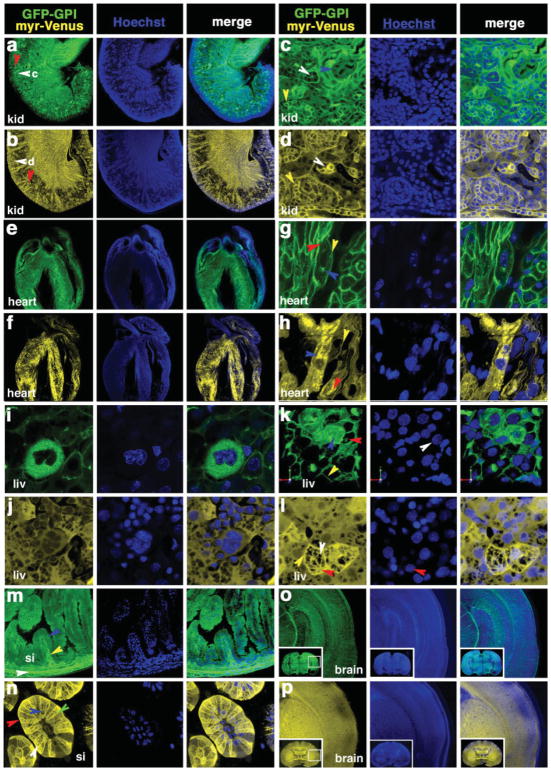
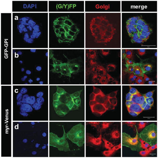
The visualization of live cell behaviors operating in situ combined with the power of mouse genetics represents a major step toward understanding the mechanisms regulating embryonic development, homeostasis, and disease progression in mammals. The availability of genetically encoded fluorescent protein reporters, combined with improved optical imaging modalities, have led to advances in our ability to examine cells in vivo. We developed a series of lipid-modified fluorescent protein fusions that are targeted to and label the secretory pathway and the plasma membrane, and that are amenable for use in mice. Here we report the generation of two strains of mice, each expressing a spectrally distinct lipid-modified GFP-variant fluorescent protein fusion. The CAG::GFP-GPI strain exhibited widespread expression of a glycosylphosphatidylinositol-tagged green fluorescent protein (GFP) fusion, while the CAG::myr-Venus strain exhibited widespread expression of a myristoyl-Venus yellow fluorescent protein fusion. Imaging of live transgenic embryonic stem (ES) cells, either live or fixed embryos and postnatal tissues demonstrated that glycosylphosphatidyl inositol- and myristoyl-tagged GFP-variant fusion proteins are targeted to and serve as markers of the plasma membrane. Moreover, our data suggest that these two lipid-modified protein fusions are dynamically targeted both to overlapping as well as distinct lipid-enriched compartments within cells. These transgenic strains not only represent high-contrast reporters of cell morphology and plasma membrane dynamics, but also may be used as in vivo sensors of lipid localization. Furthermore, combining these reporters with the study of mouse mutants will be a step forward in understanding the inter- and intracellular behaviors underlying morphogenesis in both normal and mutant contexts.
对原位发生的活细胞行为进行可视化,并结合小鼠遗传学的强大功能,是朝着理解哺乳动物胚胎发育、体内平衡和疾病进展调控机制迈出的重要一步。基因编码荧光蛋白报告基因的可用性,与改进的光学成像方式相结合,推动了我们在体内检测细胞能力的进步。我们开发了一系列脂质修饰的荧光蛋白融合体,它们靶向并标记分泌途径和质膜,且适用于小鼠。在此,我们报告了两种小鼠品系的产生,每种品系都表达一种光谱上不同的脂质修饰的绿色荧光蛋白变体荧光蛋白融合体。CAG::GFP-GPI品系表现出糖基磷脂酰肌醇标记的绿色荧光蛋白(GFP)融合体的广泛表达,而CAG::myr-Venus品系表现出肉豆蔻酰-Venus黄色荧光蛋白融合体的广泛表达。对活的转基因胚胎干细胞、活的或固定的胚胎以及出生后组织的成像表明,糖基磷脂酰肌醇和肉豆蔻酰标记的GFP变体融合蛋白靶向质膜并可作为质膜的标记物。此外,我们的数据表明,这两种脂质修饰的蛋白融合体动态靶向细胞内重叠以及不同的富含脂质的区室。这些转基因品系不仅代表了细胞形态和质膜动力学的高对比度报告基因,还可用作脂质定位的体内传感器。此外,将这些报告基因与小鼠突变体的研究相结合,将是在正常和突变背景下理解形态发生背后的细胞间和细胞内行为方面向前迈出的一步。