Artal-Sanz Marta, Samara Chrysanthi, Syntichaki Popi, Tavernarakis Nektarios
Institute of Molecular Biology and Biotechnology, Foundation for Research and Technology, Heraklion 71110, Crete, Greece.
J Cell Biol. 2006 Apr 24;173(2):231-9. doi: 10.1083/jcb.200511103.
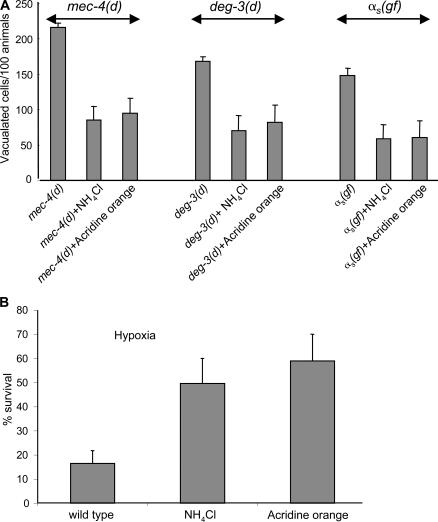
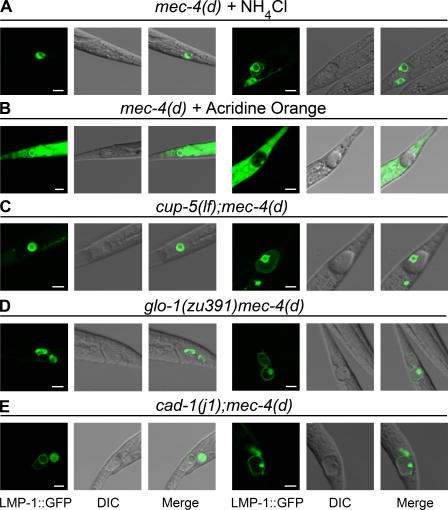
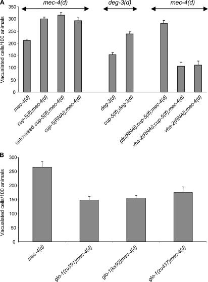
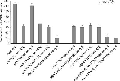
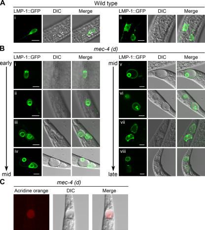
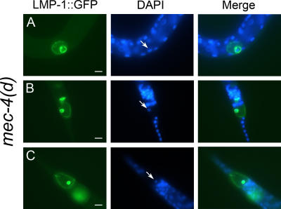
Necrotic cell death is defined by distinctive morphological characteristics that are displayed by dying cells (Walker, N.I., B.V. Harmon, G.C. Gobe, and J.F. Kerr. 1988. Methods Achiev. Exp. Pathol. 13:18-54). The cellular events that transpire during necrosis to generate these necrotic traits are poorly understood. Recent studies in the nematode Caenorhabditis elegans show that cytoplasmic acidification develops during necrosis and is required for cell death (Syntichaki, P., C. Samara, and N. Tavernarakis. 2005. Curr. Biol. 15:1249-1254). However, the origin of cytoplasmic acidification remains elusive. We show that the alkalization of endosomal and lysosomal compartments ameliorates necrotic cell death triggered by diverse stimuli. In addition, mutations in genes that result in altered lysosomal biogenesis and function markedly affect neuronal necrosis. We used a genetically encoded fluorescent marker to follow lysosome fate during neurodegeneration in vivo. Strikingly, we found that lysosomes fuse and localize exclusively around a swollen nucleus. In the advanced stages of cell death, the nucleus condenses and migrates toward the periphery of the cell, whereas green fluorescent protein-labeled lysosomal membranes fade, indicating lysosomal rupture. Our findings demonstrate a prominent role for lysosomes in cellular destruction during necrotic cell death, which is likely conserved in metazoans.
坏死性细胞死亡由垂死细胞所呈现的独特形态特征所定义(Walker, N.I., B.V. Harmon, G.C. Gobe, and J.F. Kerr. 1988. Methods Achiev. Exp. Pathol. 13:18 - 54)。在坏死过程中发生的导致这些坏死特征的细胞事件仍知之甚少。最近对线虫秀丽隐杆线虫的研究表明,坏死过程中会发生细胞质酸化,且这是细胞死亡所必需的(Syntichaki, P., C. Samara, and N. Tavernarakis. 2005. Curr. Biol. 15:1249 - 1254)。然而,细胞质酸化的起源仍然难以捉摸。我们发现,内体和溶酶体区室的碱化可改善由多种刺激引发的坏死性细胞死亡。此外,导致溶酶体生物发生和功能改变的基因突变会显著影响神经元坏死。我们使用一种基因编码的荧光标记物在体内神经退行性变过程中追踪溶酶体的命运。令人惊讶的是,我们发现溶酶体融合并仅定位在肿胀的细胞核周围。在细胞死亡的晚期,细胞核浓缩并向细胞周边迁移,而绿色荧光蛋白标记的溶酶体膜褪色,表明溶酶体破裂。我们的研究结果表明溶酶体在坏死性细胞死亡期间的细胞破坏中起重要作用,这在多细胞动物中可能是保守的。