Hsu Cheng-Da, Kymes Steven, Petrash J Mark
Department of Ophthalmology and Visual Sciences, Washington University School of Medicine, St. Louis, Missouri, USA.
Invest Ophthalmol Vis Sci. 2006 May;47(5):2036-44. doi: 10.1167/iovs.05-0524.
To characterize lenses from transgenic mice designed to express mutant and wild-type alphaA-crystallin subunits.
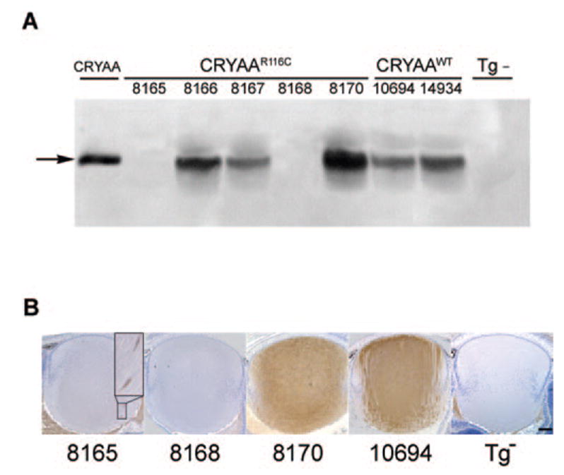
A series of transgenic mouse strains was created to express mutant (R116C) and wild-type human alphaA-crystallin in fiber cells of the lens. Dissected lenses were phenotypically scored for the presence and extent of opacities, fiber cell morphology, and posterior suture morphology. Gene transcripts derived from integrated transgenes were detected by reverse transcriptase-PCR. Distribution of expressed transgenic protein was determined by immunohistochemical staining of lens tissue sections. The abundance of endogenous and transgenic lens proteins was estimated by quantitative Western blot analysis.
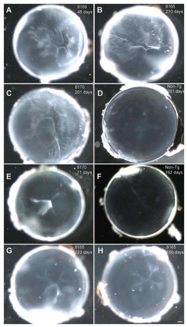
Expression of R116C mutant alphaA-crystallin subunits resulted in posterior cortical cataracts and abnormalities associated with the posterior suture. The severity of lens abnormalities did not increase between the ages of 9 and 30 weeks. With respect to opacities and morphologic abnormalities, lenses from transgenic mice that express wild-type human alphaA-crystallin subunits were indistinguishable from age-matched nontransgenic control mice. Similar phenotypes were observed in different independent lines of R116C transgenic mice that differed by at least two orders of magnitude in the expression level of the mutant transgenic protein.
The results show that lens opacities and posterior sutural defects occur when mutant R116C alphaA-crystallin subunits are expressed on the background of wild-type endogenous mouse alpha-crystallins. Low levels of R116C alphaA-crystallin subunits are sufficient to induce lens opacities and sutural defects.
对来自设计用于表达突变型和野生型αA-晶体蛋白亚基的转基因小鼠的晶状体进行特征描述。
创建了一系列转基因小鼠品系,以在晶状体的纤维细胞中表达突变型(R116C)和野生型人αA-晶体蛋白。对解剖后的晶状体进行表型评分,评估混浊的存在和程度、纤维细胞形态以及后缝合线形态。通过逆转录聚合酶链反应检测源自整合转基因的基因转录本。通过对晶状体组织切片进行免疫组织化学染色来确定表达的转基因蛋白的分布。通过定量蛋白质免疫印迹分析估计内源性和转基因晶状体蛋白的丰度。
R116C突变型αA-晶体蛋白亚基的表达导致后皮质性白内障以及与后缝合线相关的异常。在9至30周龄之间,晶状体异常的严重程度没有增加。在混浊和形态学异常方面,表达野生型人αA-晶体蛋白亚基的转基因小鼠的晶状体与年龄匹配的非转基因对照小鼠没有区别。在不同的独立R116C转基因小鼠品系中观察到了相似的表型,这些品系的突变转基因蛋白表达水平相差至少两个数量级。
结果表明,当突变型R116CαA-晶体蛋白亚基在野生型内源性小鼠α-晶体蛋白的背景下表达时,会出现晶状体混浊和后缝合线缺陷。低水平的R116CαA-晶体蛋白亚基足以诱导晶状体混浊和缝合线缺陷。