Hattar Samer, Kumar Monica, Park Alexander, Tong Patrick, Tung Jonathan, Yau King-Wai, Berson David M
Department of Neuroscience, Johns Hopkins University School of Medicine, Baltimore, Maryland 21205-2105, USA.
J Comp Neurol. 2006 Jul 20;497(3):326-49. doi: 10.1002/cne.20970.
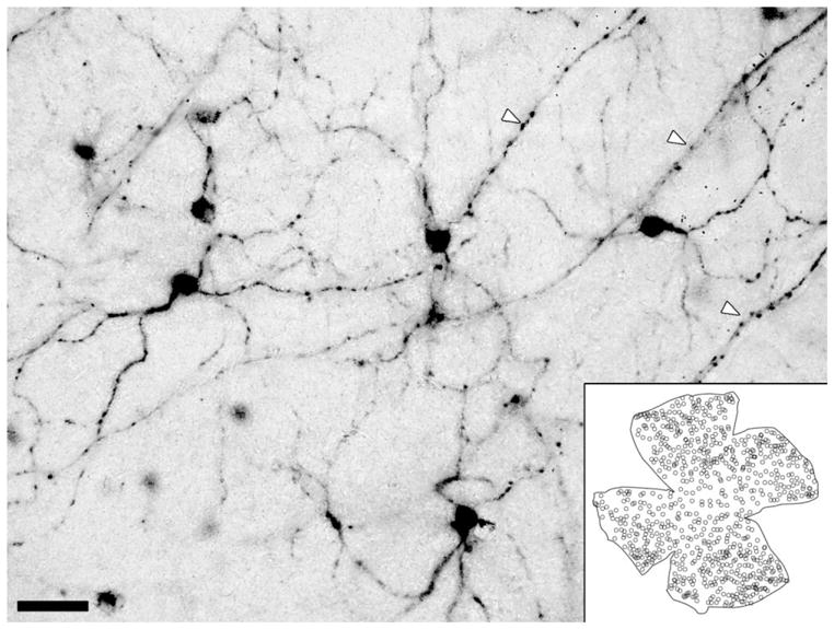
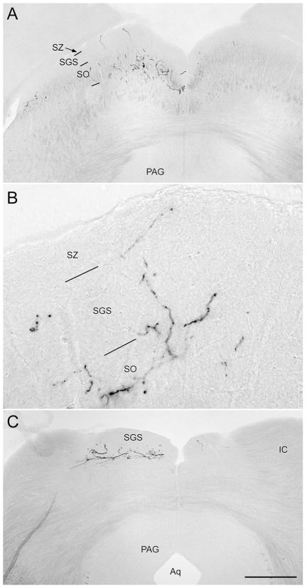
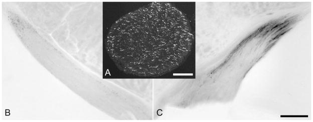
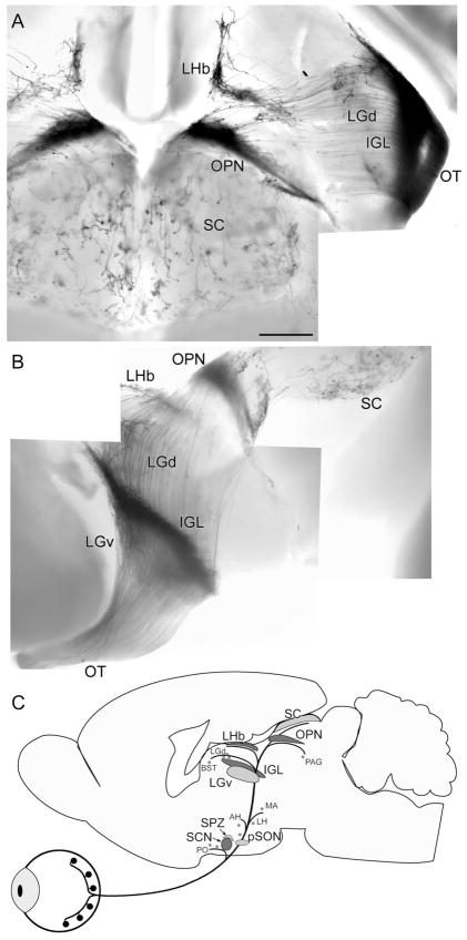
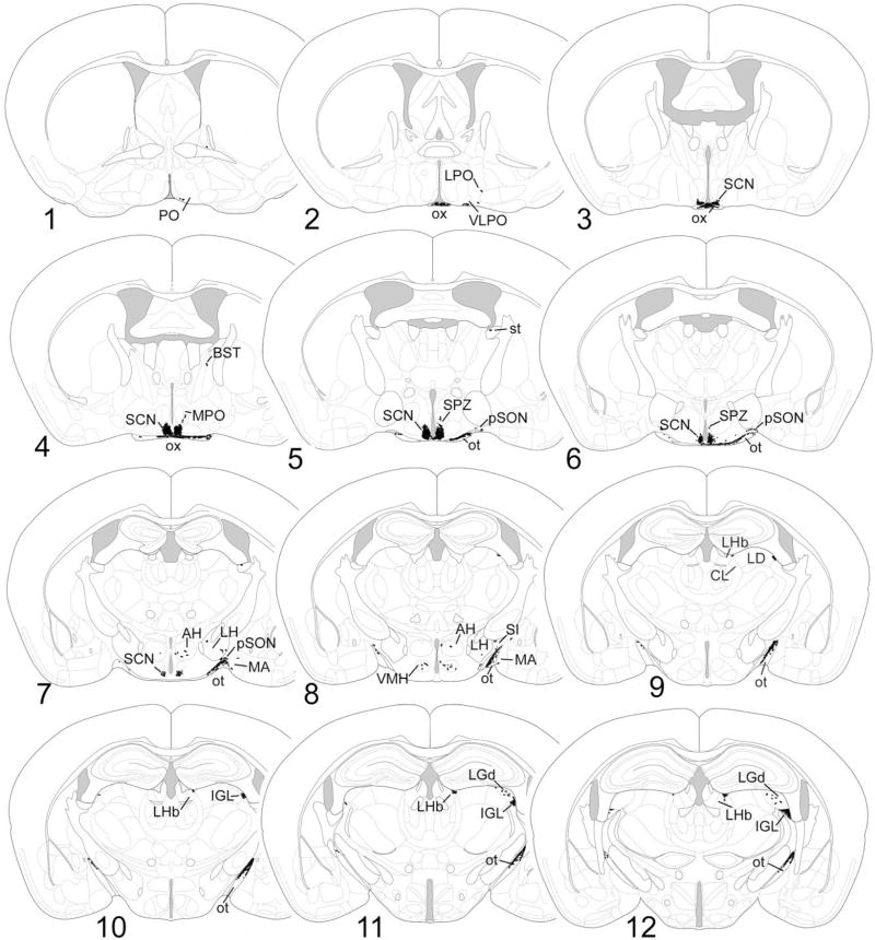
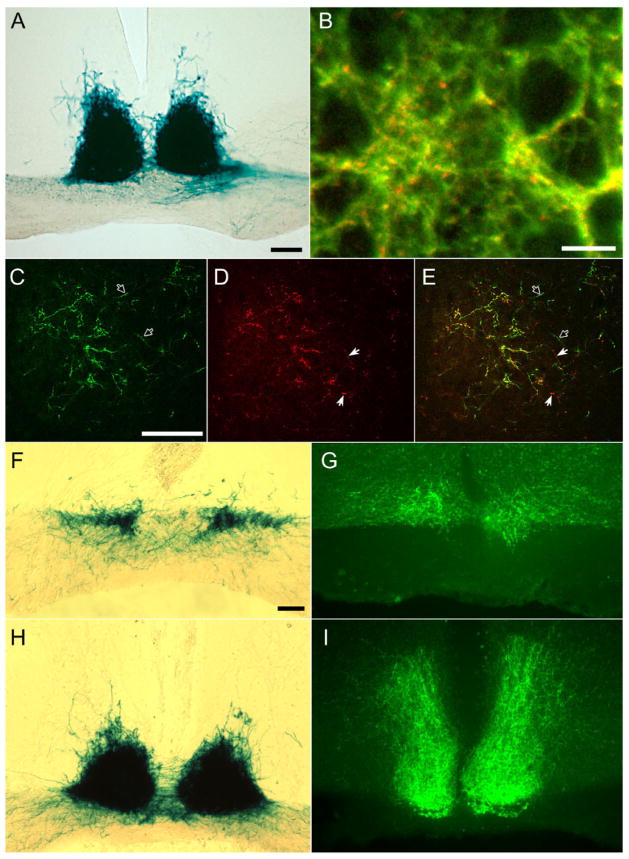
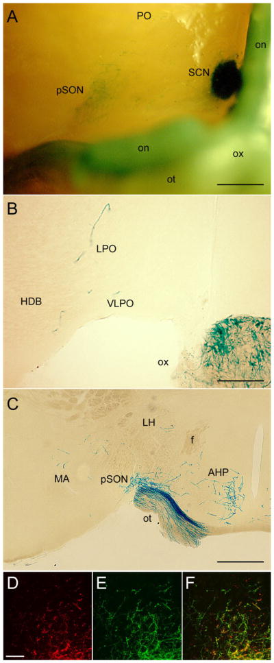
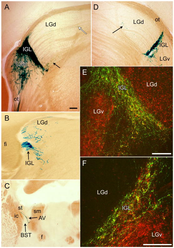
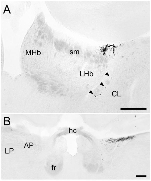
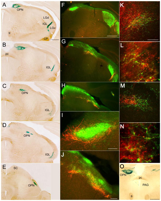
A rare type of ganglion cell in mammalian retina is directly photosensitive. These novel retinal photoreceptors express the photopigment melanopsin. They send axons directly to the suprachiasmatic nucleus (SCN), intergeniculate leaflet (IGL), and olivary pretectal nucleus (OPN), thereby contributing to photic synchronization of circadian rhythms and the pupillary light reflex. Here, we sought to characterize more fully the projections of these cells to the brain. By targeting tau-lacZ to the melanopsin gene locus in mice, ganglion cells that would normally express melanopsin were induced to express, instead, the marker enzyme beta-galactosidase. Their axons were visualized by X-gal histochemistry or anti-beta-galactosidase immunofluorescence. Established targets were confirmed, including the SCN, IGL, OPN, ventral division of the lateral geniculate nucleus (LGv), and preoptic area, but the overall projections were more widespread than previously recognized. Targets included the lateral nucleus, peri-supraoptic nucleus, and subparaventricular zone of the hypothalamus, medial amygdala, margin of the lateral habenula, posterior limitans nucleus, superior colliculus, and periaqueductal gray. There were also weak projections to the margins of the dorsal lateral geniculate nucleus. Co-staining with the cholera toxin B subunit to label all retinal afferents showed that melanopsin ganglion cells provide most of the retinal input to the SCN, IGL, and lateral habenula and much of that to the OPN, but that other ganglion cells do contribute at least some retinal input to these targets. Staining patterns after monocular enucleation revealed that the projections of these cells are overwhelmingly crossed except for the projection to the SCN, which is bilaterally symmetrical.
哺乳动物视网膜中一种罕见的神经节细胞对光直接敏感。这些新型视网膜光感受器表达光色素黑视蛋白。它们将轴突直接投射到视交叉上核(SCN)、膝间小叶(IGL)和橄榄顶盖前核(OPN),从而促进昼夜节律的光同步和瞳孔光反射。在此,我们试图更全面地描述这些细胞向大脑的投射。通过将tau - lacZ靶向小鼠的黑视蛋白基因位点,原本会表达黑视蛋白的神经节细胞被诱导转而表达标记酶β - 半乳糖苷酶。它们的轴突通过X - gal组织化学或抗β - 半乳糖苷酶免疫荧光进行可视化。确定的投射靶点得到了证实,包括SCN、IGL、OPN、外侧膝状体核腹侧部(LGv)和视前区,但总体投射比之前认为的更广泛。靶点包括下丘脑的外侧核、视上核周围核和室旁核下区、内侧杏仁核、外侧缰核边缘、后界核、上丘和导水管周围灰质。对背侧外侧膝状体核边缘也有微弱投射。用霍乱毒素B亚基共染色以标记所有视网膜传入纤维表明,黑视蛋白神经节细胞为SCN、IGL和外侧缰核提供了大部分视网膜输入,为OPN提供了许多视网膜输入,但其他神经节细胞确实至少为这些靶点贡献了一些视网膜输入。单眼摘除后的染色模式显示,除了向SCN的投射是双侧对称的外,这些细胞的投射绝大多数是交叉的。