• 文献检索
  • 文档翻译
  • 深度研究
  • 学术资讯
  • Suppr Zotero 插件Zotero 插件
  • 邀请有礼
  • 套餐&价格
  • 历史记录
应用&插件
Suppr Zotero 插件Zotero 插件浏览器插件Mac 客户端Windows 客户端微信小程序
定价
高级版会员购买积分包购买API积分包
服务
文献检索文档翻译深度研究API 文档MCP 服务
关于我们
关于 Suppr公司介绍联系我们用户协议隐私条款
关注我们

Suppr 超能文献

核心技术专利:CN118964589B侵权必究
粤ICP备2023148730 号-1Suppr @ 2026

文献检索

告别复杂PubMed语法,用中文像聊天一样搜索,搜遍4000万医学文献。AI智能推荐,让科研检索更轻松。

立即免费搜索

文件翻译

保留排版,准确专业,支持PDF/Word/PPT等文件格式,支持 12+语言互译。

免费翻译文档

深度研究

AI帮你快速写综述,25分钟生成高质量综述,智能提取关键信息,辅助科研写作。

立即免费体验

通过免疫细胞化学方法在大鼠前脑中对含磷酸酪氨酸蛋白的定位与胰岛素受体的分布相对应。

Location of phosphotyrosine-containing proteins by immunocytochemistry in the rat forebrain corresponds to the distribution of the insulin receptor.

作者信息

Moss A M, Unger J W, Moxley R T, Livingston J N

机构信息

Department of Medicine, University of Rochester School of Medicine, NY 14642.

出版信息

Proc Natl Acad Sci U S A. 1990 Jun;87(12):4453-7. doi: 10.1073/pnas.87.12.4453.

DOI:10.1073/pnas.87.12.4453
PMID:1693770
原文链接:https://pmc.ncbi.nlm.nih.gov/articles/PMC54133/
Abstract

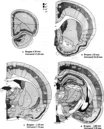
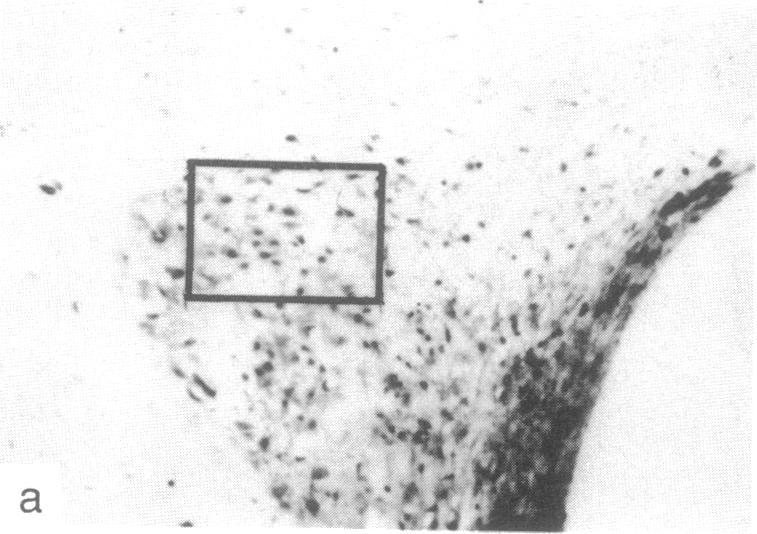
Cellular regulation by certain growth factor receptors and protooncogene products involves tyrosine kinase activity with the resultant tyrosine phosphorylation of protein substrates. In the present report we describe the distribution of phosphotyrosine-containing material detected by immunocytochemistry (ICC) in the rat forebrain. Specificity of the affinity-purified antibody against phosphotyrosine used in the ICC technique was demonstrated by the ability of phosphotyrosine and p-nitrophenyl phosphate but not phosphoserine, phosphothreonine, or L-tyrosine to inhibit the immunostaining reaction. With ICC, relatively high amounts of phosphotyrosine-positive material were observed in neurons in specific structures that included the supraoptic, paraventricular, and arcuate nuclei; the median eminence; medial habenula; subfornical organ; and piriform cortex. Moderate to high amounts were present in the cerebral cortical layers II-IV and in the pyramidal cell layer of the hippocampus. Small to moderate amounts were detected in a few other locations. Glial elements showed minimal staining. Other areas of the rat forebrain failed to react with this antibody. Importantly, the distribution of the areas positive for phosphotyrosine agreed to a remarkable extent with the distribution of the brain insulin receptor, which itself has tyrosine kinase activity. These findings suggest a relationship between the insulin receptor and the increased phosphotyrosine content of these neurons and support the concept that the brain insulin receptor is active in vivo.

摘要

某些生长因子受体和原癌基因产物对细胞的调节涉及酪氨酸激酶活性,其结果是蛋白质底物发生酪氨酸磷酸化。在本报告中,我们描述了通过免疫细胞化学(ICC)检测到的含磷酸酪氨酸物质在大鼠前脑的分布情况。ICC技术中使用的抗磷酸酪氨酸亲和纯化抗体的特异性通过以下能力得以证明:磷酸酪氨酸和对硝基苯磷酸能够抑制免疫染色反应,而磷酸丝氨酸、磷酸苏氨酸或L-酪氨酸则不能。通过ICC观察到,在特定结构的神经元中存在相对大量的磷酸酪氨酸阳性物质,这些结构包括视上核、室旁核和弓状核;正中隆起;内侧缰核;穹窿下器官;以及梨状皮质。在大脑皮质的II-IV层和海马的锥体细胞层中存在中等至大量的该物质。在其他一些位置检测到少量至中等量的该物质。神经胶质成分显示出极少的染色。大鼠前脑的其他区域与该抗体无反应。重要的是,磷酸酪氨酸阳性区域的分布在很大程度上与脑胰岛素受体的分布一致,而脑胰岛素受体本身具有酪氨酸激酶活性。这些发现表明胰岛素受体与这些神经元中磷酸酪氨酸含量的增加之间存在关联,并支持脑胰岛素受体在体内具有活性这一概念。

https://cdn.ncbi.nlm.nih.gov/pmc/blobs/db86/54133/e6258162dfea/pnas01037-0053-b.jpg
https://cdn.ncbi.nlm.nih.gov/pmc/blobs/db86/54133/e7b18fffe6af/pnas01037-0051-a.jpg
https://cdn.ncbi.nlm.nih.gov/pmc/blobs/db86/54133/44f9f08d5d4b/pnas01037-0052-a.jpg
https://cdn.ncbi.nlm.nih.gov/pmc/blobs/db86/54133/402a8e8c467e/pnas01037-0053-a.jpg
https://cdn.ncbi.nlm.nih.gov/pmc/blobs/db86/54133/e6258162dfea/pnas01037-0053-b.jpg

相似文献

1
Location of phosphotyrosine-containing proteins by immunocytochemistry in the rat forebrain corresponds to the distribution of the insulin receptor.通过免疫细胞化学方法在大鼠前脑中对含磷酸酪氨酸蛋白的定位与胰岛素受体的分布相对应。
Proc Natl Acad Sci U S A. 1990 Jun;87(12):4453-7. doi: 10.1073/pnas.87.12.4453.
2
Immunocytochemical detection of insulin receptor substrate-1 (IRS-1) in rat brain: colocalization with phosphotyrosine.大鼠脑中胰岛素受体底物-1(IRS-1)的免疫细胞化学检测:与磷酸酪氨酸的共定位
Regul Pept. 1993 Oct 20;48(1-2):257-66. doi: 10.1016/0167-0115(93)90355-c.
3
Phosphotyrosine-containing proteins in the CNS of obese Zucker rats are decreased in the absence of changes in the insulin receptor.在肥胖Zucker大鼠的中枢神经系统中,含磷酸酪氨酸的蛋白质减少,而胰岛素受体没有变化。
Neuroendocrinology. 1993 Mar;57(3):481-8. doi: 10.1159/000126395.
4
Immunohistochemical localization of insulin receptors and phosphotyrosine in the brainstem of the adult rat.成年大鼠脑干中胰岛素受体和磷酸酪氨酸的免疫组织化学定位
Neuroscience. 1991;42(3):853-61. doi: 10.1016/0306-4522(91)90049-t.
5
The hypophyseal pars tuberalis is enriched with distinct phosphotyrosine-containing proteins not detected in other areas of the brain and pituitary.垂体结节部富含在脑和垂体其他区域未检测到的独特含磷酸酪氨酸蛋白。
Cell Tissue Res. 1993 Jun;272(3):499-507. doi: 10.1007/BF00318556.
6
Distribution of insulin receptor-like immunoreactivity in the rat forebrain.胰岛素受体样免疫反应性在大鼠前脑的分布。
Neuroscience. 1989;31(1):143-57. doi: 10.1016/0306-4522(89)90036-5.
7
Insulin stimulates the tyrosine phosphorylation of a 61-kilodalton protein in rat adipocytes.胰岛素刺激大鼠脂肪细胞中一种61千道尔顿蛋白质的酪氨酸磷酸化。
Endocrinology. 1992 Mar;130(3):1533-8. doi: 10.1210/endo.130.3.1371454.
8
In the absence of antibody IgGsorb precipitates human placenta phosphotyrosine-containing proteins.在没有抗体的情况下,IgGsorb沉淀人胎盘含磷酸酪氨酸的蛋白质。
Biochem Biophys Res Commun. 1989 Feb 28;159(1):332-6. doi: 10.1016/0006-291x(89)92442-x.
9
Analysis of insulin receptor phosphorylation sites in intact rat liver cells by two-dimensional phosphopeptide mapping. Predominance of the tris-phosphorylated form of the kinase domain after stimulation by insulin.通过二维磷酸肽图谱分析完整大鼠肝细胞中的胰岛素受体磷酸化位点。胰岛素刺激后激酶结构域的三磷酸化形式占主导地位。
Biochem J. 1991 Apr 1;275 ( Pt 1)(Pt 1):15-21. doi: 10.1042/bj2750015.
10
Distinct beta-subunits are present in hybrid insulin-like-growth-factor-1 receptors in the central nervous system.不同的β亚基存在于中枢神经系统的杂合胰岛素样生长因子-1受体中。
Biochem J. 1993 Sep 15;294 ( Pt 3)(Pt 3):685-92. doi: 10.1042/bj2940685.

引用本文的文献

1
Cellular Profile of Subfornical Organ Insulin Receptors in Mice.SUBFORNICAL ORGAN 胰岛素受体在小鼠中的细胞特征。
Biomolecules. 2024 Oct 4;14(10):1256. doi: 10.3390/biom14101256.
2
State of the Science on Brain Insulin Resistance and Cognitive Decline Due to Alzheimer's Disease.脑胰岛素抵抗与阿尔茨海默病导致的认知衰退的科学现状
Aging Dis. 2024 Aug 1;15(4):1688-1725. doi: 10.14336/AD.2023.0814.
3
Cerebrovascular insulin receptors are defective in Alzheimer's disease.阿尔茨海默病患者的脑血管胰岛素受体存在缺陷。

本文引用的文献

1
The insulin receptor of rat brain is coupled to tyrosine kinase activity.大鼠大脑的胰岛素受体与酪氨酸激酶活性相关联。
J Biol Chem. 1984 Mar 25;259(6):3470-4.
2
Regional distribution of calcium- and cyclic adenosine 3':5'-monophosphate-regulated protein phosphorylation systems in mammalian brain. II. Soluble systems.哺乳动物脑中钙和环磷酸腺苷调节的蛋白质磷酸化系统的区域分布。II. 可溶性系统。
J Neurosci. 1983 Feb;3(2):302-11. doi: 10.1523/JNEUROSCI.03-02-00302.1983.
3
pp60c-src in the developing cerebellum.发育中小脑的pp60c-src
Brain. 2023 Jan 5;146(1):75-90. doi: 10.1093/brain/awac309.
4
Exercise with 40-Hz light flicker improves hippocampal insulin signaling in Alzheimer disease mice.40赫兹光闪烁运动可改善阿尔茨海默病小鼠的海马胰岛素信号传导。
J Exerc Rehabil. 2022 Feb 24;18(1):20-27. doi: 10.12965/jer.2244042.021. eCollection 2022 Feb.
5
Subfornical organ insulin receptors tonically modulate cardiovascular and metabolic function.穹窿下器官胰岛素受体持续调节心血管和代谢功能。
Physiol Genomics. 2019 Aug 1;51(8):333-341. doi: 10.1152/physiolgenomics.00021.2019. Epub 2019 Jun 7.
6
The role of hypothalamic mTORC1 signaling in insulin regulation of food intake, body weight, and sympathetic nerve activity in male mice.下丘脑mTORC1信号通路在雄性小鼠胰岛素调节食物摄入、体重和交感神经活动中的作用。
Endocrinology. 2015 Apr;156(4):1398-407. doi: 10.1210/en.2014-1660. Epub 2015 Jan 9.
7
Deregulation of brain insulin signaling in Alzheimer's disease.阿尔茨海默病中大脑胰岛素信号传导的失调。
Neurosci Bull. 2014 Apr;30(2):282-94. doi: 10.1007/s12264-013-1408-x. Epub 2014 Mar 20.
8
Insulin in the brain: sources, localization and functions.脑内胰岛素:来源、定位和功能。
Mol Neurobiol. 2013 Feb;47(1):145-71. doi: 10.1007/s12035-012-8339-9. Epub 2012 Sep 7.
9
Effects of age on elements of insulin-signaling pathway in central nervous system of rats.年龄对大鼠中枢神经系统胰岛素信号通路元件的影响。
Endocrine. 2001 Dec;16(3):227-34. doi: 10.1385/endo:16:3:227.
10
The hypophyseal pars tuberalis is enriched with distinct phosphotyrosine-containing proteins not detected in other areas of the brain and pituitary.垂体结节部富含在脑和垂体其他区域未检测到的独特含磷酸酪氨酸蛋白。
Cell Tissue Res. 1993 Jun;272(3):499-507. doi: 10.1007/BF00318556.
Mol Cell Biol. 1985 Jan;5(1):27-32. doi: 10.1128/mcb.5.1.27-32.1985.
4
Localization and characterization of insulin receptors in rat brain and pituitary gland using in vitro autoradiography and computerized densitometry.利用体外放射自显影和计算机化密度测定法对大鼠脑和垂体中的胰岛素受体进行定位和特性分析。
Endocrinology. 1987 Oct;121(4):1562-70. doi: 10.1210/endo-121-4-1562.
5
Insulin in the brain.大脑中的胰岛素。
Annu Rev Physiol. 1987;49:335-47. doi: 10.1146/annurev.ph.49.030187.002003.
6
Localization of 125I-insulin binding sites in the rat hypothalamus by quantitative autoradiography.通过定量放射自显影法对大鼠下丘脑125I-胰岛素结合位点进行定位。
Neurosci Lett. 1986 Sep 25;70(1):17-22. doi: 10.1016/0304-3940(86)90430-1.
7
An early developmental phase of pp60c-src expression in the neural ectoderm.神经外胚层中pp60c-src表达的早期发育阶段。
Dev Biol. 1986 Sep;117(1):83-9. doi: 10.1016/0012-1606(86)90350-7.
8
Autoradiographic localization of insulin receptors in rat brain: prominence in olfactory and limbic areas.大鼠脑中胰岛素受体的放射自显影定位:在嗅觉和边缘系统区域较为突出。
Neuroscience. 1986 Apr;17(4):1127-38. doi: 10.1016/0306-4522(86)90082-5.
9
Localization of binding sites for insulin-like growth factor-I (IGF-I) in the rat brain by quantitative autoradiography.通过定量放射自显影法对大鼠脑中胰岛素样生长因子-I(IGF-I)结合位点进行定位。
Brain Res. 1988 Mar 22;444(2):205-13. doi: 10.1016/0006-8993(88)90931-6.
10
Immunocytochemical localization of protein kinase C in developing brain tissue and in primary neuronal cultures.蛋白激酶C在发育中的脑组织和原代神经元培养物中的免疫细胞化学定位。
Dev Biol. 1988 Mar;126(1):98-107. doi: 10.1016/0012-1606(88)90243-6.