Grivas Theodoros B, Burwell Geoffrey R, Vasiliadis Elias S, Webb John K
Orthopaedic Department, Thriasion General Hospital, G. Gennimata Av. 19600, Magoula, Attica, Greece.
Scoliosis. 2006 Oct 18;1:17. doi: 10.1186/1748-7161-1-17.
The role of rib cage in the development of progressive infantile idiopathic scoliosis (IIS) has not been studied previously. No report was found for rib growth in children with IIS. These findings caused us to undertake a segmental radiological study of the spine and rib-cage in children with progressive IIS. The aim of the present study is to present a new method for assessing the thoracic shape in scoliotics and in control subjects and to compare the findings between the two groups.
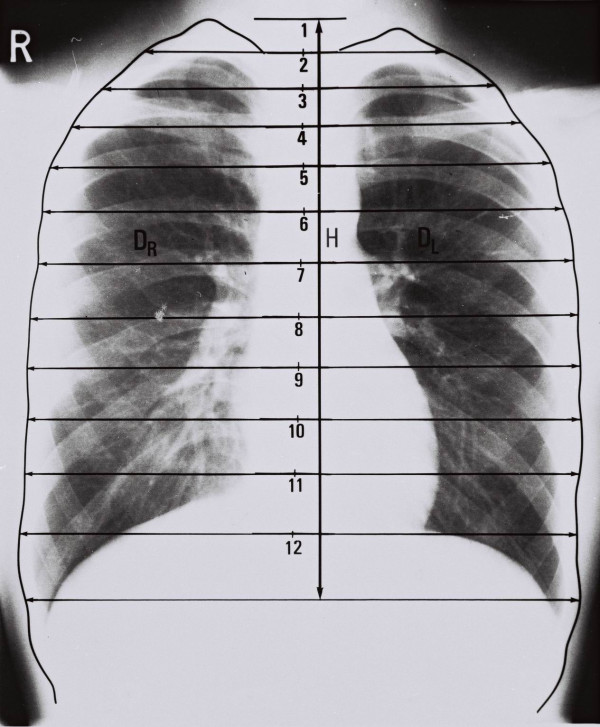
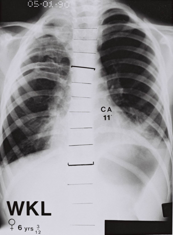
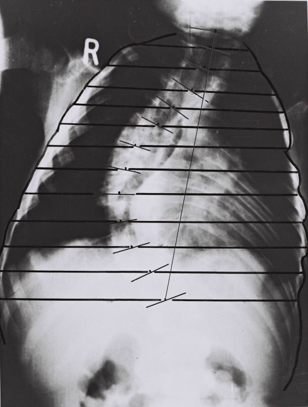
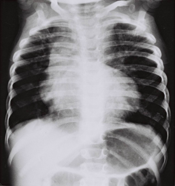
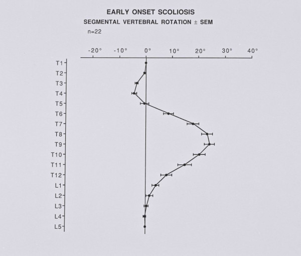
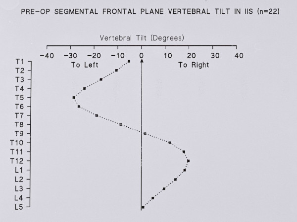
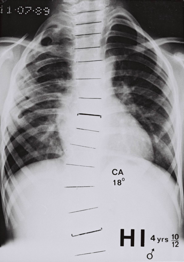
In the posteroanterior (PA) spinal radiographs of 24 patients with progressive IIS, with a mean age of 4.1 years old, the Thoracic Ratios (TRs) (segmental convex and concave TRs), the Cobb angle, the segmental vertebral rotation and vertebral tilt were measured. In 233 subjects, with a mean age of 5.1 years old, who were used as a control group, the segmental left and right TRs and the total width of the chest (left plus right TRs) were measured in PA chest radiographs. Statistical analysis included Mann-Whitney, Spearman correlation coefficient, multiple linear regression analysis and ANOVA.
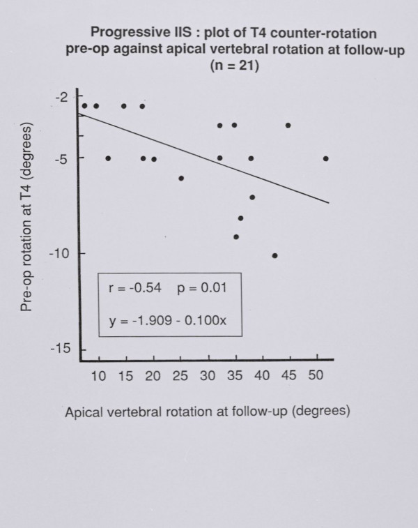
The comparison shows that the scoliotic thorax is significantly narrower than that of the controls at all spinal levels. The upper chest in IIS is funnel-shaped and the vertebral rotation at T4 early in management correlates significantly with the apical vertebral rotation at follow up.
The IIS thorax is narrower than that of the controls, the upper chest is funnel-shaped and there is a predictive value of vertebral rotation at the upper limit of the thoracic curve of IIS, which reflects, impaired rib control of spinal rotation possibly due to neuromuscular factors, which contribute also to the funnel-shaped chest.
胸廓在进行性婴儿特发性脊柱侧凸(IIS)发展过程中的作用此前尚未得到研究。未发现有关IIS患儿肋骨生长的报告。这些发现促使我们对进行性IIS患儿的脊柱和胸廓进行分段放射学研究。本研究的目的是提出一种评估脊柱侧凸患者和对照受试者胸廓形状的新方法,并比较两组的研究结果。
在24例平均年龄4.1岁的进行性IIS患者的后前位(PA)脊柱X线片上,测量胸廓比率(TRs)(节段性凸侧和凹侧TRs)、Cobb角、节段性椎体旋转和椎体倾斜度。在233例平均年龄5.1岁的作为对照组的受试者的PA胸部X线片上,测量节段性左右TRs和胸廓总宽度(左右TRs之和)。统计分析包括Mann-Whitney检验、Spearman相关系数、多元线性回归分析和方差分析。
比较显示,在所有脊柱节段,脊柱侧凸患者的胸廓明显比对照组窄。IIS患者的上胸部呈漏斗状,治疗早期T4椎体的旋转与随访时顶椎的旋转显著相关。
IIS患者的胸廓比对照组窄,上胸部呈漏斗状,IIS胸弯上限处椎体旋转具有预测价值,这反映出可能由于神经肌肉因素导致肋骨对脊柱旋转的控制受损,这也导致了漏斗状胸部的形成。