McKinley M P, Meyer R K, Kenaga L, Rahbar F, Cotter R, Serban A, Prusiner S B
Department of Neurology, University of California, San Francisco 94143.
J Virol. 1991 Mar;65(3):1340-51. doi: 10.1128/JVI.65.3.1340-1351.1991.
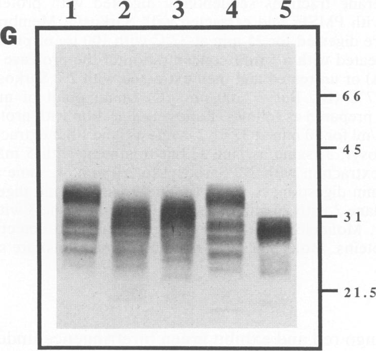
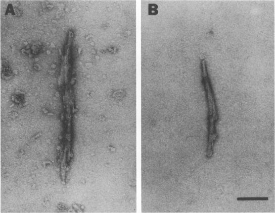
Scrapie prion infectivity can be enriched from hamster brain homogenates by using limited proteolysis and detergent extraction. Purified fractions contain both scrapie infectivity and the protein PrP 27-30, which is aggregated in the form of prion rods. During purification, PrP 27-30 is produced from a larger membrane protein, PrPSc, by limited proteolysis with proteinase K. Brain homogenates from scrapie-infected hamsters do not contain prion rods prior to exposure to detergents and proteases. To determine whether both detergent extraction and limited proteolysis are required for the formation of prion rods, microsomal membranes were prepared from infected brains in the presence of protease inhibitors. The isolated membranes were then detergent extracted as well as protease digested to evaluate the effects of these treatments on the formation of prion rods. Neither detergent (2% Sarkosyl) extraction nor limited proteinase K digestion of scrapie microsomes produced recognizable prion amyloid rods. Only after combining detergent extraction with limited proteolysis were numerous prion rods observed. Rod formation was influenced by the protease concentration, the specificity of the protease, and the duration of digestion. Rod formation also depended upon the detergent; some combinations of protease and detergent did not produce prion amyloid rods. Similar results were obtained with purified PrPSc fractions prepared by repeated detergent extractions in the presence of protease inhibitors. These fractions contained amorphous structures but not rods; however, prion rods were produced upon conversion of PrPSc to PrP 27-30 by limited proteolysis. We conclude that the formation of prion amyloid rods in vitro requires both detergent extraction and limited proteolysis. In vivo, amyloid filaments found in the brains of animals with scrapie resemble prion rods in their width and their labeling with prion protein (PrP) antisera; however, filaments are typically longer than rods. Whether limited proteolysis and some process equivalent to detergent extraction are required for amyloid filament formation in vivo remains to be established.
通过有限蛋白酶解和去污剂提取,可从仓鼠脑匀浆中富集瘙痒病朊病毒感染性。纯化后的组分既含有瘙痒病感染性,也含有以朊病毒杆状形式聚集的蛋白质PrP 27-30。在纯化过程中,PrP 27-30是由一种更大的膜蛋白PrPSc经蛋白酶K有限蛋白酶解产生的。来自感染瘙痒病仓鼠的脑匀浆在未接触去污剂和蛋白酶之前不含朊病毒杆。为了确定形成朊病毒杆是否需要去污剂提取和有限蛋白酶解,在蛋白酶抑制剂存在的情况下从感染的脑中制备微粒体膜。然后对分离的膜进行去污剂提取以及蛋白酶消化,以评估这些处理对朊病毒杆形成的影响。瘙痒病微粒体的去污剂(2%十二烷基肌氨酸钠)提取或有限的蛋白酶K消化均未产生可识别的朊病毒淀粉样杆。只有在将去污剂提取与有限蛋白酶解相结合后,才观察到大量的朊病毒杆。杆的形成受蛋白酶浓度、蛋白酶的特异性以及消化持续时间的影响。杆的形成还取决于去污剂;某些蛋白酶和去污剂的组合不会产生朊病毒淀粉样杆。用在蛋白酶抑制剂存在下通过反复去污剂提取制备的纯化PrPSc组分也得到了类似的结果。这些组分含有无定形结构但不含杆;然而,通过有限蛋白酶解将PrPSc转化为PrP 27-30后产生了朊病毒杆。我们得出结论,体外朊病毒淀粉样杆的形成需要去污剂提取和有限蛋白酶解。在体内,患有瘙痒病的动物脑中发现的淀粉样细丝在宽度和用朊病毒蛋白(PrP)抗血清标记方面类似于朊病毒杆;然而,细丝通常比杆长。体内淀粉样细丝形成是否需要有限蛋白酶解和某种等同于去污剂提取的过程仍有待确定。