Kelsen Jesper, Kjaer Katrine, Chen Gang, Pedersen Michael, Røhl Lisbeth, Frøkiaer Jørgen, Nielsen Søren, Nyengaard Jens R, Rønn Lars Christian B
The Water and Salt Research Centre, University of Aarhus, DK-8000 Aarhus C, Denmark.
J Neuroinflammation. 2006 Dec 6;3:31. doi: 10.1186/1742-2094-3-31.
Anti-inflammatory treatment affects ischemic damage and neurogenesis in rodent models of cerebral ischemia. We investigated the potential benefit of COX-2 inhibition with parecoxib in spontaneously hypertensive rats (SHRs) subjected to transient middle cerebral artery occlusion (tMCAo).
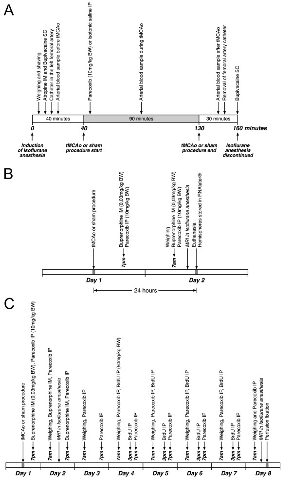
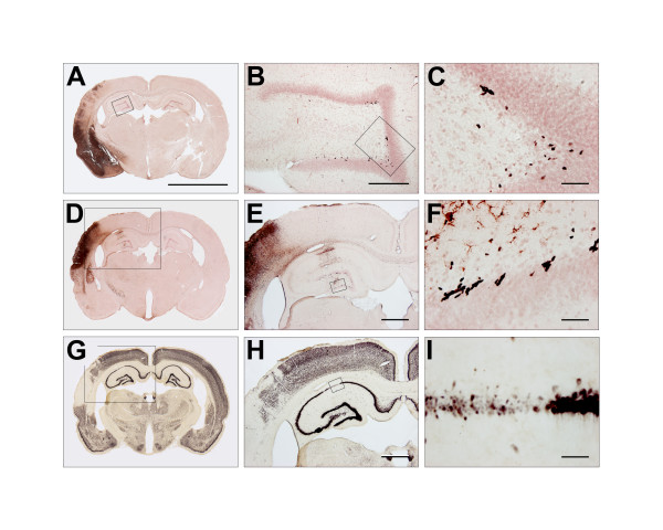
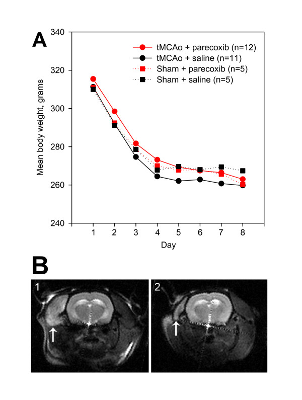
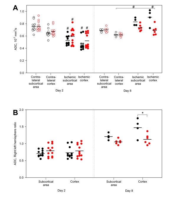
Sixty-four male SHRs were randomized to 90 min of intraluminal tMCAo or sham surgery. Parecoxib (10 mg/kg) or isotonic saline was administered intraperitoneally (IP) during the procedure, and twice daily thereafter. Nineteen animals were euthanized after 24 hours, and each hemisphere was examined for mRNA expression of pro-inflammatory cytokines and COX enzymes by quantitative RT-PCR. Twenty-three tMCAo animals were studied with diffusion and T2 weighted MRI within the first 24 hours, and ten of the SHRs underwent follow-up MRI six days later. Thirty-three SHRs were given 5-bromo-2'-deoxy-uridine (BrdU) twice daily on Day 4 to 7 after tMCAo. Animals were euthanized on Day 8 and the brains were studied with free-floating immunohistochemistry for activated microglia (ED-1), hippocampal granule cell BrdU incorporation, and neuronal nuclei (NeuN). Infarct volume estimation was done using the 2D nucleator and Cavalieri principle on NeuN-stained coronal brain sections. The total number of BrdU+ cells in the dentate gyrus (DG) of the hippocampus was estimated using the optical fractionator.
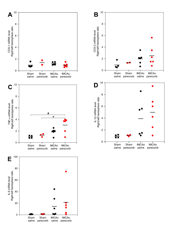
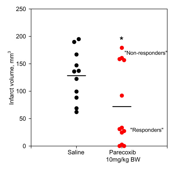
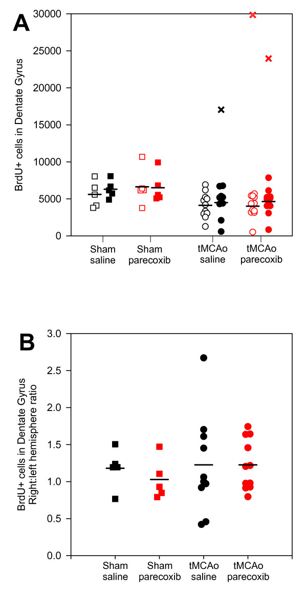
We found a significant reduction in infarct volume in parecoxib treated animals one week after tMCAo (p < 0.03). Cortical ADC values in the parecoxib group were markedly less increased on Day 8 (p < 0.01). Interestingly, the parecoxib treated rats were segregated into two subgroups, suggesting a responder vs. non-responder phenomenon. We found indications of mRNA up-regulation of IL-1beta, IL-6, TNF-alpha and COX-2, whereas COX-1 remained unaffected. Hippocampal granule cell BrdU incorporation was not affected by parecoxib treatment. Presence of ED-1+ activated microglia in the hippocampus was related to an increase in BrdU uptake in the DG.
IP parecoxib administration during tMCAo was neuroprotective, as evidenced by a large reduction in mean infarct volume and a lower cortical ADC increment. Increased pro-inflammatory cytokine mRNA levels and hippocampal granule cell BrdU incorporation remained unaffected.
在脑缺血的啮齿动物模型中,抗炎治疗会影响缺血性损伤和神经发生。我们研究了帕瑞昔布抑制环氧化酶-2(COX-2)对短暂大脑中动脉闭塞(tMCAo)的自发性高血压大鼠(SHR)的潜在益处。
64只雄性SHR被随机分为接受90分钟管腔内tMCAo或假手术。在手术过程中腹腔注射(IP)帕瑞昔布(10mg/kg)或等渗盐水,此后每天两次。24小时后对19只动物实施安乐死,通过定量逆转录聚合酶链反应(RT-PCR)检测每个半球促炎细胞因子和COX酶的mRNA表达。在最初24小时内对23只tMCAo动物进行扩散加权和T2加权磁共振成像(MRI)检查,其中10只SHR在6天后接受随访MRI检查。在tMCAo后第4至7天,对33只SHR每天两次给予5-溴-2'-脱氧尿苷(BrdU)。在第8天对动物实施安乐死,通过游离漂浮免疫组织化学法研究大脑中活化小胶质细胞(ED-1)、海马颗粒细胞BrdU掺入情况以及神经元核(NeuN)。使用二维核计数器和卡瓦列里原理在NeuN染色的冠状脑切片上估计梗死体积。使用光学分割器估计海马齿状回(DG)中BrdU+细胞的总数。
我们发现tMCAo一周后,帕瑞昔布治疗组动物的梗死体积显著减小(p<0.03)。在第8天,帕瑞昔布组的皮质表观扩散系数(ADC)值增加明显较少(p<−0.01)。有趣的是,帕瑞昔布治疗的大鼠被分为两个亚组,提示存在反应者与无反应者现象。我们发现白细胞介素-1β(IL-1β)、白细胞介素-6(IL-6)、肿瘤坏死因子-α(TNF-α)和COX-2的mRNA上调,而COX-1不受影响。帕瑞昔布治疗不影响海马颗粒细胞BrdU掺入。海马中ED-1+活化小胶质细胞的存在与DG中BrdU摄取增加有关。
tMCAo期间腹腔注射帕瑞昔布具有神经保护作用,平均梗死体积大幅减小和皮质ADC增量较低证明了这一点。促炎细胞因子mRNA水平升高和海马颗粒细胞BrdU掺入不受影响。