Poser Benedikt A, Norris David G
FC Donders Centre for Cognitive Neuroimaging, Nijmegen, The Netherlands.
MAGMA. 2007 Apr;20(2):63-7. doi: 10.1007/s10334-007-0068-0. Epub 2007 Feb 21.
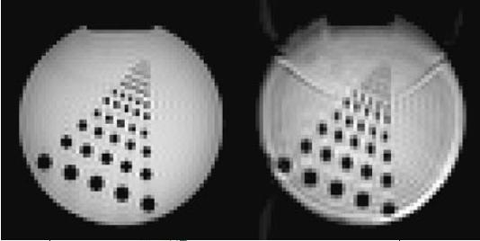
The recently developed vascular space occupancy (VASO) fMRI technique is gaining popularity as it facilitates the measurement of cerebral blood volume (CBV) changes concomitant with brain activation, without the use of contrast agents. Thus far, VASO fMRI has only been used in conjunction with a GE-EPI (gradient-echo echo planar imaging) sequence, which is proceeded by an inversion recovery (IR) experiment to selectively null the blood signal. The use of GE-EPI has potential disadvantages: (a) the non-zero TE may lead to BOLD contamination and (b) images suffer from the EPI-typical inhomogeneity artefacts.
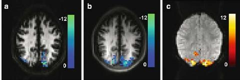
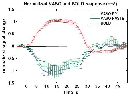
Here, we propose the use of VASO based on an IR-HASTE (inversion recovery half-Fourier acquisition single-shot turbo spin echo) sequence. Results Results from a visual stimulation study (n = 8) show a 43% higher functional contrast-to-noise (CNR) of HASTE compared to EPI, with a strongly increased count of active voxels at the same significance threshold. Sensitivity to inflow effects was investigated and found to be similar for both methods.
As HASTE VASO yields essentially artefact-free images, it appears to be the method of choice for measuring relative CBV changes with VASO.
最近开发的血管空间占据(VASO)功能磁共振成像技术正越来越受欢迎,因为它有助于在不使用造影剂的情况下测量与脑激活相关的脑血容量(CBV)变化。到目前为止,VASO功能磁共振成像仅与GE-EPI(梯度回波平面回波成像)序列结合使用,该序列之前进行反转恢复(IR)实验以选择性地消除血液信号。GE-EPI的使用存在潜在缺点:(a)非零回波时间(TE)可能导致血氧水平依赖(BOLD)信号污染,(b)图像存在典型的EPI不均匀伪影。
在此,我们提出基于IR-HASTE(反转恢复半傅里叶采集单次激发快速自旋回波)序列使用VASO。结果一项视觉刺激研究(n = 8)的结果表明,与EPI相比,HASTE的功能对比噪声比(CNR)高43%,在相同显著性阈值下,激活体素数量大幅增加。对流入效应的敏感性进行了研究,发现两种方法相似。
由于HASTE VASO产生的图像基本无伪影,它似乎是用VASO测量相对CBV变化的首选方法。