Toni Mattia, Spisni Enzo, Griffoni Cristiana, Santi Spartaco, Riccio Massimo, Lenaz Patrizia, Tomasi Vittorio
Department of Experimental Biology, University of Bologna, Bologna, Italy.
J Biomed Biotechnol. 2006;2006(5):69469. doi: 10.1155/JBB/2006/69469.
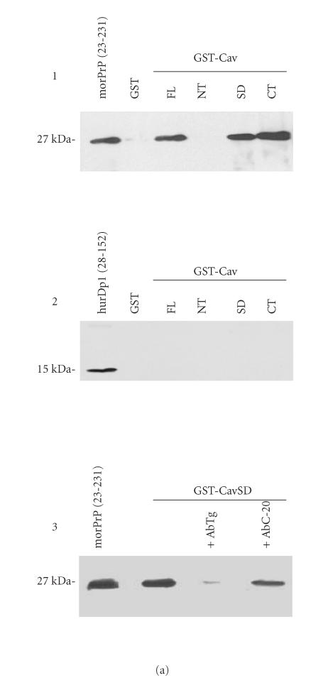
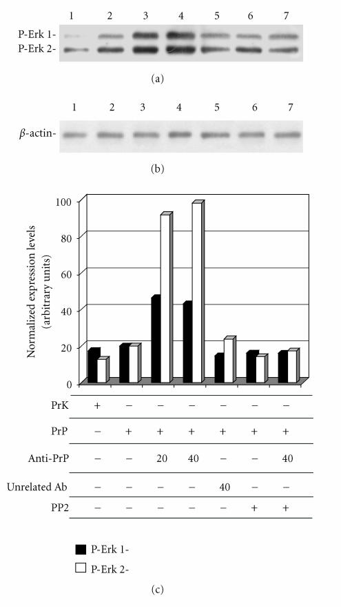
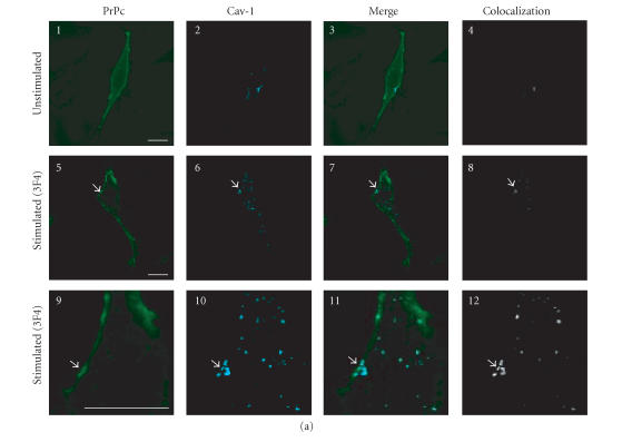
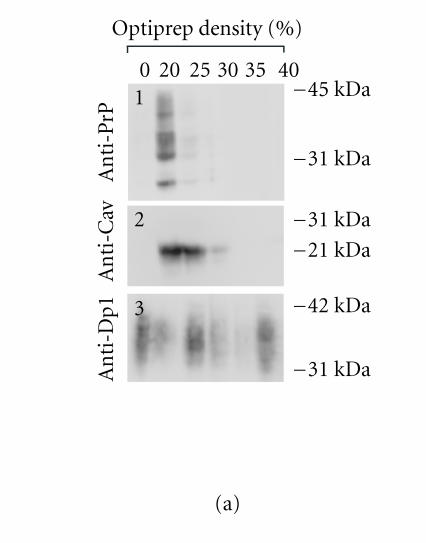
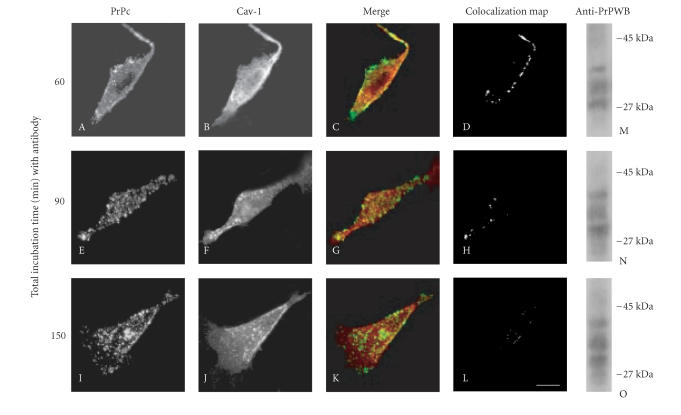
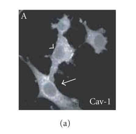
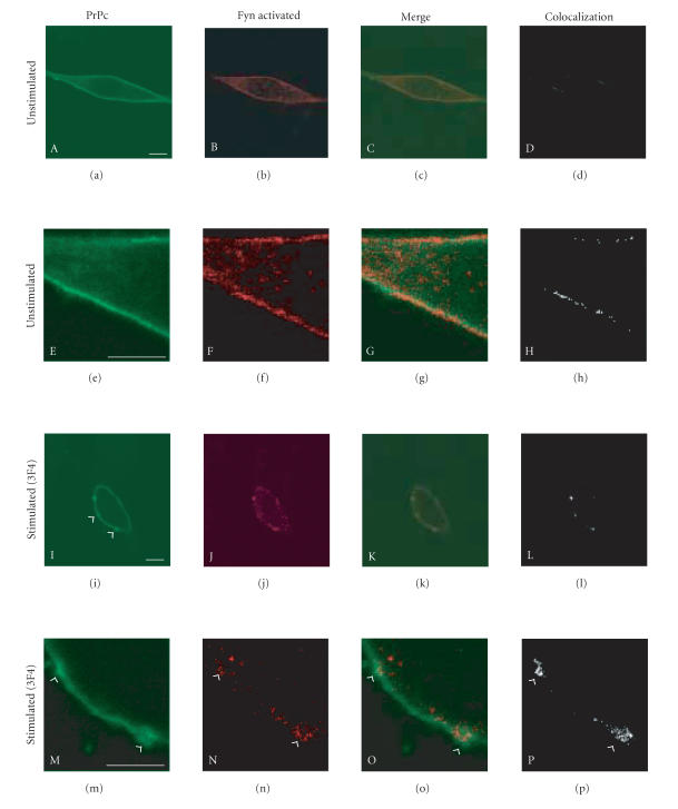
It has been reported that cellular prion protein (PrPc) is enriched in caveolae or caveolae-like domains with caveolin-1 (Cav-1) participating to signal transduction events by Fyn kinase recruitment. By using the Glutathione-S-transferase (GST)-fusion proteins assay, we observed that PrPc strongly interacts in vitro with Cav-1. Thus, we ascertained the PrPc caveolar localization in a hypothalamic neuronal cell line (GN11), by confocal microscopy analysis, flotation on density gradient, and coimmunoprecipitation experiments. Following the anti-PrPc antibody-mediated stimulation of live GN11 cells, we observed that PrPc clustered on plasma membrane domains rich in Cav-1 in which Fyn kinase converged to be activated. After these events, a signaling cascade through p42/44 MAP kinase (Erk 1/2) was triggered, suggesting that following translocations from rafts to caveolae or caveolae-like domains PrPc could interact with Cav-1 and induce signal transduction events.
据报道,细胞朊蛋白(PrPc)在小窝或类小窝结构域中富集,小窝蛋白-1(Cav-1)通过招募Fyn激酶参与信号转导事件。通过使用谷胱甘肽-S-转移酶(GST)融合蛋白分析,我们观察到PrPc在体外与Cav-1强烈相互作用。因此,我们通过共聚焦显微镜分析、密度梯度浮选和免疫共沉淀实验,确定了PrPc在下丘脑神经元细胞系(GN11)中的小窝定位。在用抗PrPc抗体介导刺激活的GN11细胞后,我们观察到PrPc聚集在富含Cav-1的质膜结构域上,Fyn激酶在其中聚集并被激活。在这些事件之后,通过p42/44丝裂原活化蛋白激酶(Erk 1/2)触发了信号级联反应,这表明从脂筏转移到小窝或类小窝结构域后,PrPc可能与Cav-1相互作用并诱导信号转导事件。