Hooks Darren A
Bioengineering Institute, University of Auckland, New Zealand.
Biomed Eng Online. 2007 Jun 5;6:21. doi: 10.1186/1475-925X-6-21.
Computer models of the electrical and mechanical actions of the heart, solved on geometrically realistic domains, are becoming an increasingly useful scientific tool. Construction of these models requires detailed measurement of the microstructural features which impact on the function of the heart. Currently a few generic cardiac models are in use for a wide range of simulation problems, and contributions to publicly accessible databases of cardiac structures, on which models can be solved, remain rare. This paper presents to-date the largest database of porcine left ventricular segment microstructural architecture, for use in both electrical and mechanical simulation.
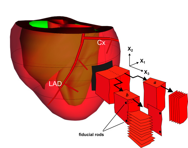
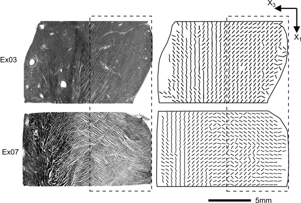
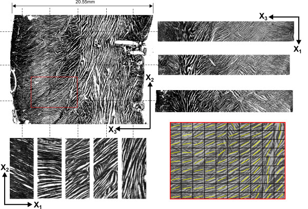
Cryosectioning techniques were used to reconstruct the myofibre and myosheet orientations in tissue blocks of size ~15 x 15 x 15 mm, taken from the mid-anterior left ventricular freewall, of seven hearts. Tissue sections were gathered on orthogonal planes, and the angles of intersection of myofibres and myosheets with these planes determined automatically with a gradient intensity based algorithm. These angles were then combined to provide a description of myofibre and myosheet variation throughout the tissue, in a form able to be input to biophysically based computational models of the heart.
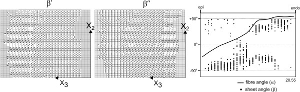
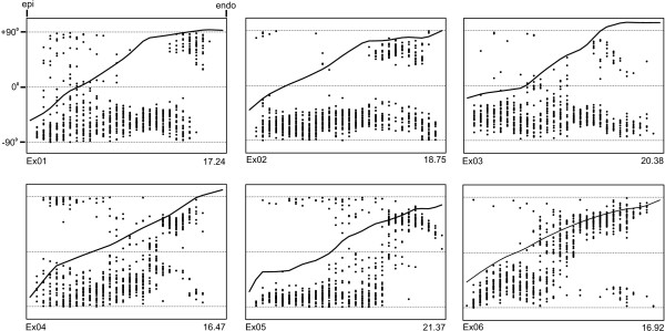
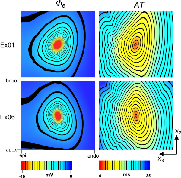
Several microstructural features were common across all hearts. Myofibres rotated through 141 +/- 18 degrees (mean +/- SD) from epicardium to endocardium, in near linear fashion. In the outer two-thirds of the wall sheet angles were predominantly negative, however, in the inner one-third an abrupt change in sheet angle, with reversal in sign, was seen in six of the seven hearts. Two distinct populations of sheets with orthogonal orientations often co-existed, usually with one population dominating. The utility of the tissue structures was demonstrated by simulating the passive and active electrical responses of two of the tissue blocks to current injection. Distinct patterns of electrical response were obtained in the two tissue blocks, illustrating the importance of testing model based predictions on a variety of tissue architectures.
This study significantly expands the set of geometries on which models of cardiac function can be solved.
在几何逼真的区域上求解的心脏电活动和机械活动的计算机模型正日益成为一种有用的科学工具。构建这些模型需要对影响心脏功能的微观结构特征进行详细测量。目前,一些通用的心脏模型被用于广泛的模拟问题,而对可供公开访问的心脏结构数据库(可在其上求解模型)的贡献仍然很少。本文展示了迄今为止最大的猪左心室节段微观结构数据库,可用于电模拟和机械模拟。
采用冷冻切片技术重建取自七颗心脏左心室前壁中部、大小约为15×15×15mm组织块中的肌纤维和肌片方向。在正交平面上收集组织切片,并使用基于梯度强度的算法自动确定肌纤维和肌片与这些平面的相交角度。然后将这些角度组合起来,以一种能够输入到基于生物物理学的心脏计算模型的形式,描述整个组织中肌纤维和肌片的变化。
所有心脏都有几个共同的微观结构特征。肌纤维从心外膜到心内膜以近似线性的方式旋转了141±18度(平均值±标准差)。在心室壁外三分之二部分,肌片角度主要为负,然而,在七颗心脏中的六颗心脏中,在内三分之一处观察到肌片角度突然变化且符号反转。通常存在两个具有正交方向的不同肌片群体,通常其中一个群体占主导。通过模拟其中两个组织块对电流注入的被动和主动电反应,证明了组织结构的实用性。在两个组织块中获得了不同的电反应模式,说明了在各种组织结构上测试基于模型的预测的重要性。
本研究显著扩展了可用于求解心脏功能模型的几何形状集合。