• 文献检索
  • 文档翻译
  • 深度研究
  • 学术资讯
  • Suppr Zotero 插件Zotero 插件
  • 邀请有礼
  • 套餐&价格
  • 历史记录
应用&插件
Suppr Zotero 插件Zotero 插件浏览器插件Mac 客户端Windows 客户端微信小程序
定价
高级版会员购买积分包购买API积分包
服务
文献检索文档翻译深度研究API 文档MCP 服务
关于我们
关于 Suppr公司介绍联系我们用户协议隐私条款
关注我们

Suppr 超能文献

核心技术专利:CN118964589B侵权必究
粤ICP备2023148730 号-1Suppr @ 2026

文献检索

告别复杂PubMed语法,用中文像聊天一样搜索,搜遍4000万医学文献。AI智能推荐,让科研检索更轻松。

立即免费搜索

文件翻译

保留排版,准确专业,支持PDF/Word/PPT等文件格式,支持 12+语言互译。

免费翻译文档

深度研究

AI帮你快速写综述,25分钟生成高质量综述,智能提取关键信息,辅助科研写作。

立即免费体验

光学生物传感器可区分A431细胞中内源性PAR1和PAR2的信号传导。

Optical biosensor differentiates signaling of endogenous PAR1 and PAR2 in A431 cells.

作者信息

Fang Ye, Ferrie Ann M

机构信息

Biochemical Technologies, Science and Technology Division, Corning Incorporated, Sullivan Park, Corning, NY 14831, USA.

出版信息

BMC Cell Biol. 2007 Jun 22;8:24. doi: 10.1186/1471-2121-8-24.

DOI:10.1186/1471-2121-8-24
PMID:17587449
原文链接:https://pmc.ncbi.nlm.nih.gov/articles/PMC1925066/
Abstract

BACKGROUND

Protease activated receptors (PARs) consist of a family of four G protein-coupled receptors. Many types of cells express several PARs, whose physiological significance is mostly unknown.

RESULTS

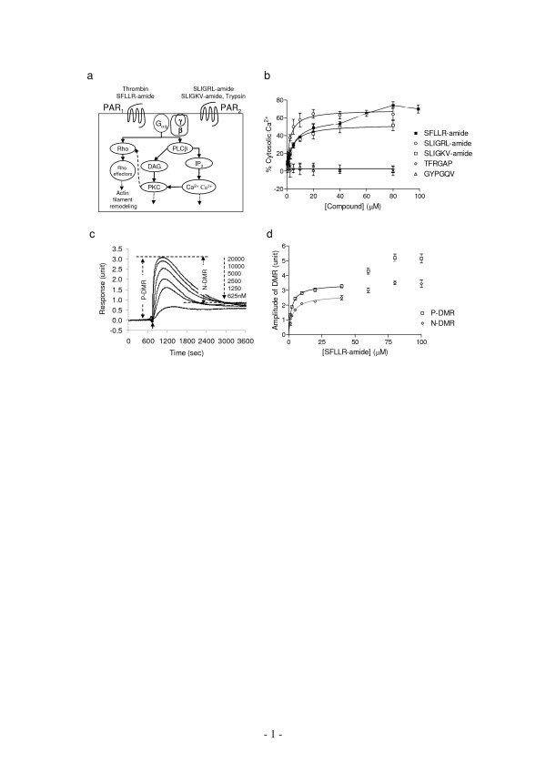
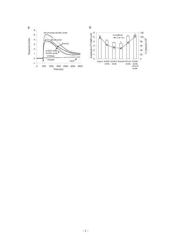
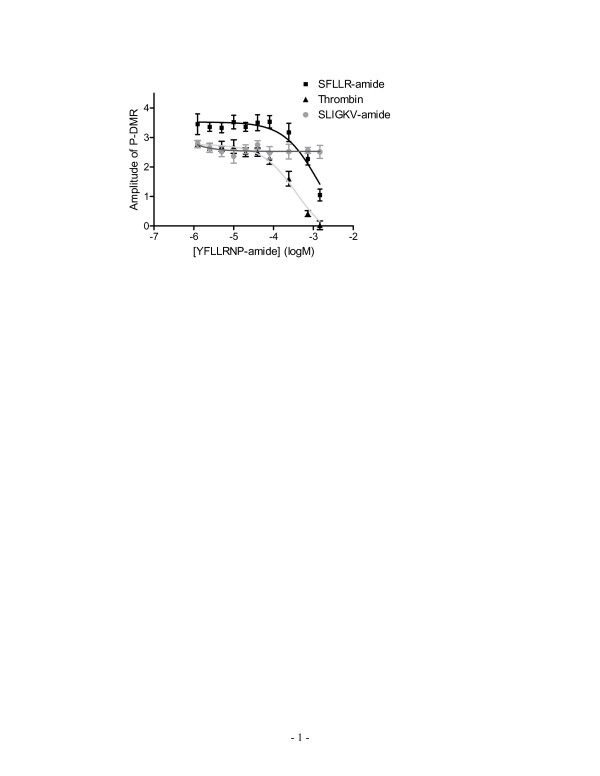
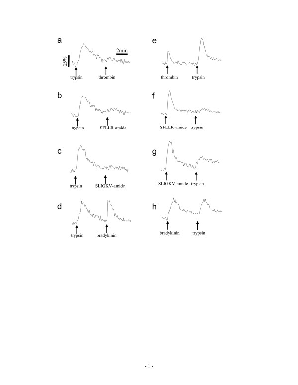
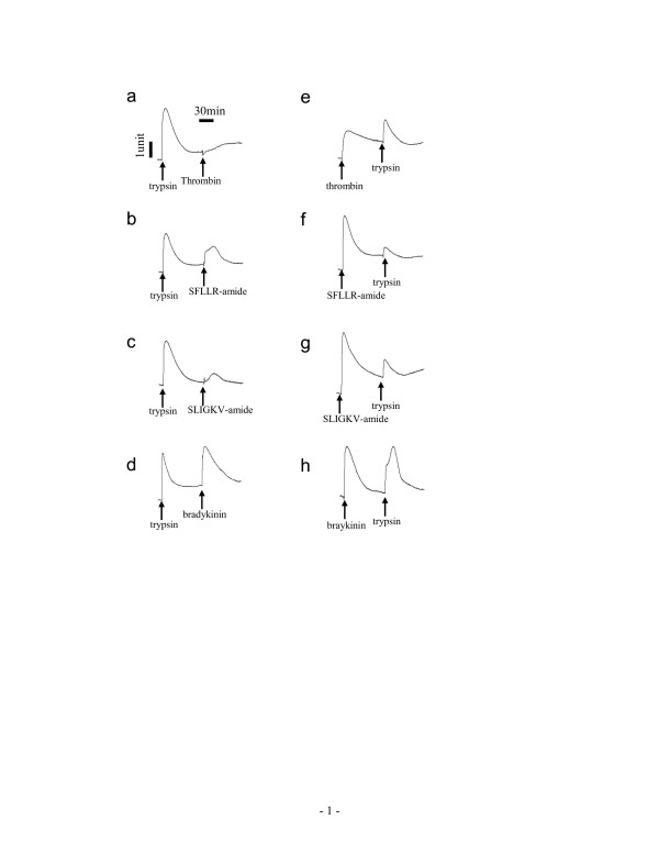
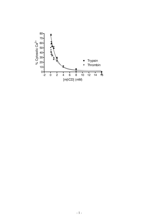
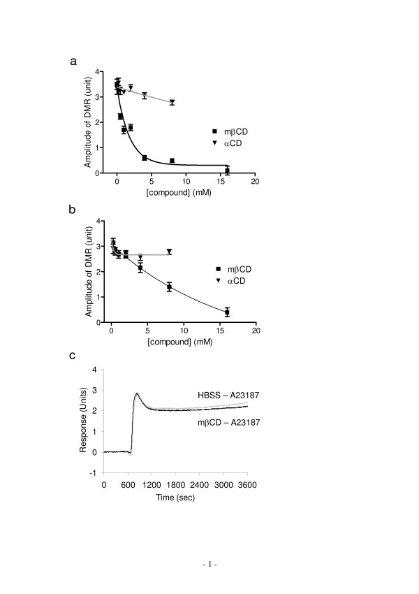
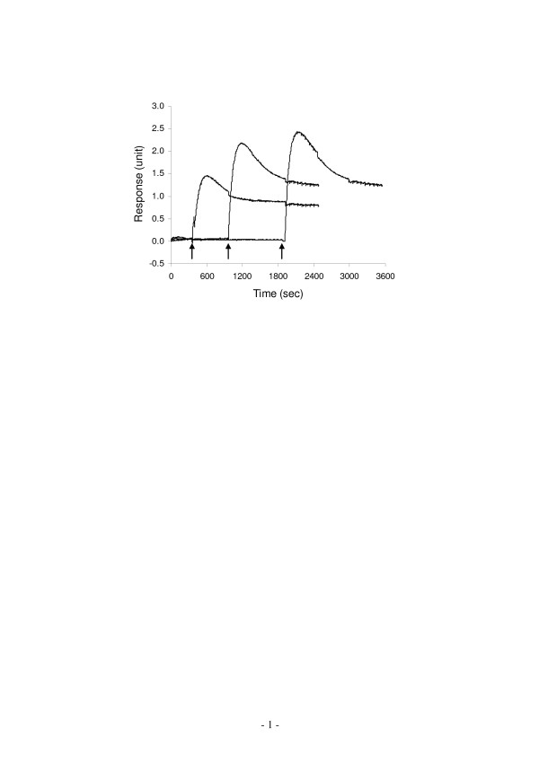
Here, we show that non-invasive resonant waveguide grating (RWG) biosensor differentiates signaling of endogenous protease activated receptor subtype 1 (PAR1) and 2 (PAR2) in human epidermoid carcinoma A431 cells. The biosensor directly measures dynamic mass redistribution (DMR) resulted from ligand-induced receptor activation in adherent cells. In A431, both PAR1 and PAR2 agonists, but neither PAR3 nor PAR4 agonists, trigger dose-dependent Ca2+ mobilization as well as Gq-type DMR signals. Both Ca2+ flux and DMR signals display comparable desensitization patterns upon repeated stimulation with different combinations of agonists. However, PAR1 and PAR2 exhibit distinct kinetics of receptor re-sensitization. Furthermore, both trypsin- and thrombin-induced Ca2+ flux signals show almost identical dependence on cell surface cholesterol level, but their corresponding DMR signals present different sensitivities.

CONCLUSION

Optical biosensor provides an alternative readout for examining receptor activation under physiologically relevant conditions, and differentiates the signaling of endogenous PAR1 and PAR2 in A431.

摘要

背景

蛋白酶激活受体(PARs)由四种G蛋白偶联受体组成的家族。许多类型的细胞表达几种PARs,其生理意义大多未知。

结果

在此,我们表明非侵入性共振波导光栅(RWG)生物传感器可区分人表皮样癌A431细胞中内源性蛋白酶激活受体亚型1(PAR1)和2(PAR2)的信号传导。该生物传感器直接测量贴壁细胞中配体诱导的受体激活所导致的动态质量再分布(DMR)。在A431细胞中,PAR1和PAR2激动剂均可触发剂量依赖性的Ca2+动员以及Gq型DMR信号,而PAR3和PAR4激动剂则不能。在用不同组合的激动剂重复刺激后,Ca2+通量和DMR信号均显示出可比的脱敏模式。然而,PAR1和PAR2表现出不同的受体再敏化动力学。此外,胰蛋白酶和凝血酶诱导的Ca2+通量信号对细胞表面胆固醇水平的依赖性几乎相同,但其相应的DMR信号表现出不同的敏感性。

结论

光学生物传感器为在生理相关条件下检测受体激活提供了一种替代读数,并区分了A431细胞中内源性PAR1和PAR2的信号传导。

https://cdn.ncbi.nlm.nih.gov/pmc/blobs/df35/1925066/24cff164ee2e/1471-2121-8-24-9.jpg
https://cdn.ncbi.nlm.nih.gov/pmc/blobs/df35/1925066/925ca36f8174/1471-2121-8-24-1.jpg
https://cdn.ncbi.nlm.nih.gov/pmc/blobs/df35/1925066/516db63a3a84/1471-2121-8-24-2.jpg
https://cdn.ncbi.nlm.nih.gov/pmc/blobs/df35/1925066/aec302d1768a/1471-2121-8-24-3.jpg
https://cdn.ncbi.nlm.nih.gov/pmc/blobs/df35/1925066/a7ad8fb4ff8d/1471-2121-8-24-4.jpg
https://cdn.ncbi.nlm.nih.gov/pmc/blobs/df35/1925066/226d49d58485/1471-2121-8-24-5.jpg
https://cdn.ncbi.nlm.nih.gov/pmc/blobs/df35/1925066/28d5e2cc647d/1471-2121-8-24-6.jpg
https://cdn.ncbi.nlm.nih.gov/pmc/blobs/df35/1925066/08b3995e4d5f/1471-2121-8-24-7.jpg
https://cdn.ncbi.nlm.nih.gov/pmc/blobs/df35/1925066/9eef029f404e/1471-2121-8-24-8.jpg
https://cdn.ncbi.nlm.nih.gov/pmc/blobs/df35/1925066/24cff164ee2e/1471-2121-8-24-9.jpg

相似文献

1
Optical biosensor differentiates signaling of endogenous PAR1 and PAR2 in A431 cells.光学生物传感器可区分A431细胞中内源性PAR1和PAR2的信号传导。
BMC Cell Biol. 2007 Jun 22;8:24. doi: 10.1186/1471-2121-8-24.
2
Protease-activated receptors (PAR1 and PAR2) contribute to tumor cell motility and metastasis.蛋白酶激活受体(PAR1和PAR2)有助于肿瘤细胞的运动和转移。
Mol Cancer Res. 2004 Jul;2(7):395-402.
3
Protease-activated receptor-2 is essential for factor VIIa and Xa-induced signaling, migration, and invasion of breast cancer cells.蛋白酶激活受体-2对于因子VIIa和Xa诱导的乳腺癌细胞信号传导、迁移及侵袭至关重要。
Cancer Res. 2006 Jan 1;66(1):307-14. doi: 10.1158/0008-5472.CAN-05-1735.
4
Protease-activated receptor mediated RhoA signaling and cytoskeletal reorganization in LNCaP cells.蛋白酶激活受体介导的LNCaP细胞中的RhoA信号传导和细胞骨架重组。
Biochemistry. 2003 Jan 28;42(3):702-9. doi: 10.1021/bi027100x.
5
Prostate-specific kallikreins-2 and -4 enhance the proliferation of DU-145 prostate cancer cells through protease-activated receptors-1 and -2.前列腺特异性激肽释放酶-2和-4通过蛋白酶激活受体-1和-2促进DU-145前列腺癌细胞的增殖。
Mol Cancer Res. 2008 Jun;6(6):1043-51. doi: 10.1158/1541-7786.MCR-08-0096.
6
Activated protein C signals through the thrombin receptor PAR1 in endothelial cells.活化蛋白C通过内皮细胞中的凝血酶受体PAR1发出信号。
J Endotoxin Res. 2003;9(5):317-21. doi: 10.1179/096805103225002584.
7
Evaluation of proteinase-activated receptor-1 (PAR1) agonists and antagonists using a cultured cell receptor desensitization assay: activation of PAR2 by PAR1-targeted ligands.使用培养细胞受体脱敏试验评估蛋白酶激活受体-1(PAR1)激动剂和拮抗剂:PAR1靶向配体对PAR2的激活作用
J Pharmacol Exp Ther. 1999 Jan;288(1):358-70.
8
Proteinase-activated receptors 1 and 2 in rat olfactory system: layer-specific regulation of multiple signaling pathways in the main olfactory bulb and induction of neurite retraction in olfactory sensory neurons.大鼠嗅觉系统中的蛋白酶激活受体1和2:主嗅球中多种信号通路的层特异性调节以及嗅觉感觉神经元中神经突回缩的诱导。
Neuroscience. 2007 May 25;146(3):1289-301. doi: 10.1016/j.neuroscience.2007.02.059. Epub 2007 Apr 16.
9
Inflammatory mediators modulate thrombin and cathepsin-G signaling in human bronchial fibroblasts by inducing expression of proteinase-activated receptor-4.炎症介质通过诱导蛋白酶激活受体-4的表达来调节人支气管成纤维细胞中的凝血酶和组织蛋白酶G信号传导。
Am J Physiol Lung Cell Mol Physiol. 2007 Mar;292(3):L788-98. doi: 10.1152/ajplung.00226.2006. Epub 2006 Dec 1.
10
Optical biosensor provides insights for bradykinin B(2) receptor signaling in A431 cells.光学生物传感器为A431细胞中缓激肽B(2)受体信号传导提供了见解。
FEBS Lett. 2005 Nov 21;579(28):6365-74. doi: 10.1016/j.febslet.2005.10.019. Epub 2005 Oct 25.

引用本文的文献

1
Metasurface-enhanced infrared spectroscopy in multiwell format for real-time assaying of live cells.多井格式的亚表面增强红外光谱法用于实时分析活细胞。
Lab Chip. 2023 May 2;23(9):2228-2240. doi: 10.1039/d3lc00017f.
2
Probing the Drug Dynamics of Chemotherapeutics Using Metasurface-Enhanced Infrared Reflection Spectroscopy of Live Cells.利用活细胞的亚表面增强红外反射光谱技术探测化疗药物动力学。
Cells. 2022 May 10;11(10):1600. doi: 10.3390/cells11101600.
3
Monitoring the effects of chemical stimuli on live cells with metasurface-enhanced infrared reflection spectroscopy.

本文引用的文献

1
Non-invasive optical biosensor for assaying endogenous G protein-coupled receptors in adherent cells.用于检测贴壁细胞内源性G蛋白偶联受体的非侵入式光学生物传感器。
J Pharmacol Toxicol Methods. 2007 May-Jun;55(3):314-22. doi: 10.1016/j.vascn.2006.11.001. Epub 2006 Nov 29.
2
Dosage-dependent switch from G protein-coupled to G protein-independent signaling by a GPCR.G蛋白偶联受体(GPCR)介导的剂量依赖性从G蛋白偶联信号传导切换为非G蛋白依赖性信号传导
EMBO J. 2007 Jan 10;26(1):53-64. doi: 10.1038/sj.emboj.7601502. Epub 2006 Dec 14.
3
Resonant waveguide grating biosensor for living cell sensing.
利用超表面增强红外反射光谱监测活细胞中化学刺激的影响。
Lab Chip. 2021 Oct 12;21(20):3991-4004. doi: 10.1039/d1lc00580d.
4
Pharmacological profile of the neuropeptide S receptor: Dynamic mass redistribution studies.神经肽 S 受体的药理学特征:动态质量重分布研究。
Pharmacol Res Perspect. 2018 Dec 3;6(6):e00445. doi: 10.1002/prp2.445. eCollection 2018 Dec.
5
NOP receptor pharmacological profile - A dynamic mass redistribution study.NOP 受体药理学特征——一项动态质量重分布研究。
PLoS One. 2018 Aug 30;13(8):e0203021. doi: 10.1371/journal.pone.0203021. eCollection 2018.
6
Overexpression of Na/Mg exchanger SLC41A1 attenuates pro-survival signaling.钠/镁交换体SLC41A1的过表达减弱了促生存信号传导。
Oncotarget. 2017 Dec 22;9(4):5084-5104. doi: 10.18632/oncotarget.23598. eCollection 2018 Jan 12.
7
Non-invasive Optical Biosensor for Probing Cell Signaling.用于探测细胞信号传导的非侵入式光学生物传感器
Sensors (Basel). 2007 Oct 16;7(10):2316-2329. doi: 10.3390/s7102316.
8
Recent Advances in Biosensing With Photonic Crystal Surfaces: A Review.光子晶体表面生物传感的最新进展:综述
IEEE Sens J. 2016 May 15;16(10):3349-3366. doi: 10.1109/JSEN.2015.2429738. Epub 2015 May 5.
9
Intensity interrogation near cutoff resonance for label-free cellular profiling.用于无标记细胞分析的截止共振附近的强度询问
Sci Rep. 2016 Apr 18;6:24685. doi: 10.1038/srep24685.
10
Microplate-compatible total internal reflection fluorescence microscopy for receptor pharmacology.用于受体药理学的微孔板兼容全内反射荧光显微镜
Appl Phys Lett. 2013 May 13;102(19):193702. doi: 10.1063/1.4805041. Epub 2013 May 14.
用于活细胞传感的共振波导光栅生物传感器。
Biophys J. 2006 Sep 1;91(5):1925-40. doi: 10.1529/biophysj.105.077818. Epub 2006 Jun 9.
4
Cellular functions of cholesterol probed with optical biosensors.利用光学生物传感器探究胆固醇的细胞功能。
Biochim Biophys Acta. 2006 Feb;1763(2):254-61. doi: 10.1016/j.bbamcr.2006.01.006. Epub 2006 Feb 13.
5
Optical biosensor provides insights for bradykinin B(2) receptor signaling in A431 cells.光学生物传感器为A431细胞中缓激肽B(2)受体信号传导提供了见解。
FEBS Lett. 2005 Nov 21;579(28):6365-74. doi: 10.1016/j.febslet.2005.10.019. Epub 2005 Oct 25.
6
Characteristics of dynamic mass redistribution of epidermal growth factor receptor signaling in living cells measured with label-free optical biosensors.使用无标记光学生物传感器测量活细胞中表皮生长因子受体信号动态质量再分布的特征
Anal Chem. 2005 Sep 1;77(17):5720-5. doi: 10.1021/ac050887n.
7
Functional selectivity of G protein signaling by agonist peptides and thrombin for the protease-activated receptor-1.激动剂肽和凝血酶对蛋白酶激活受体-1的G蛋白信号传导功能选择性
J Biol Chem. 2005 Jul 1;280(26):25048-59. doi: 10.1074/jbc.M414090200. Epub 2005 May 4.
8
Binding of a highly potent protease-activated receptor-2 (PAR2) activating peptide, [3H]2-furoyl-LIGRL-NH2, to human PAR2.一种高效的蛋白酶激活受体-2(PAR2)激活肽[3H]2-呋喃甲酰-LIGRL-NH2与人PAR2的结合。
Br J Pharmacol. 2005 May;145(2):255-63. doi: 10.1038/sj.bjp.0706189.
9
Translocation of diacylglycerol kinase theta from cytosol to plasma membrane in response to activation of G protein-coupled receptors and protein kinase C.响应G蛋白偶联受体和蛋白激酶C的激活,二酰基甘油激酶θ从胞质溶胶转位至质膜。
J Biol Chem. 2005 Mar 18;280(11):9870-8. doi: 10.1074/jbc.M409301200. Epub 2005 Jan 4.
10
Involvement of G protein betagamma-subunits in diverse signaling induced by G(i/o)-coupled receptors: study using the Xenopus oocyte expression system.G蛋白βγ亚基参与G(i/o)偶联受体诱导的多种信号传导:利用非洲爪蟾卵母细胞表达系统进行的研究
Am J Physiol Cell Physiol. 2004 Oct;287(4):C885-94. doi: 10.1152/ajpcell.00125.2004. Epub 2004 May 19.