Hao Ying, Sood Sumita, Triadafilopoulos George, Kim Jong Hyeok, Wang Zheng, Sahbaie Peyman, Omary M Bishr, Lowe Anson W
Department of Medicine, Stanford University, Stanford, CA, USA.
BMC Gastroenterol. 2007 Jun 27;7:24. doi: 10.1186/1471-230X-7-24.
Esophageal reflux and Barrett's esophagus represent two major risk factors for the development of esophageal adenocarcinoma. Previous studies have shown that brief exposure of the Barrett's-associated adenocarcinoma cell line, SEG-1, or primary cultures of Barrett's esophageal tissues to acid or bile results in changes consistent with cell proliferation. In this study, we determined whether similar exposure to acid or bile salts results in gene expression changes that provide insights into malignant transformation.
Using previously published methods, Barrett's-associated esophageal adenocarcinoma cell lines and primary cultures of Barrett's esophageal tissue were exposed to short pulses of acid or bile salts followed by incubation in culture media at pH 7.4. A genome-wide assessment of gene expression was then determined for the samples using cDNA microarrays. Subsequent analysis evaluated for statistical differences in gene expression with and without treatment.
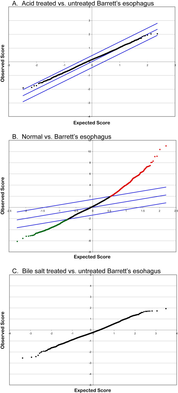
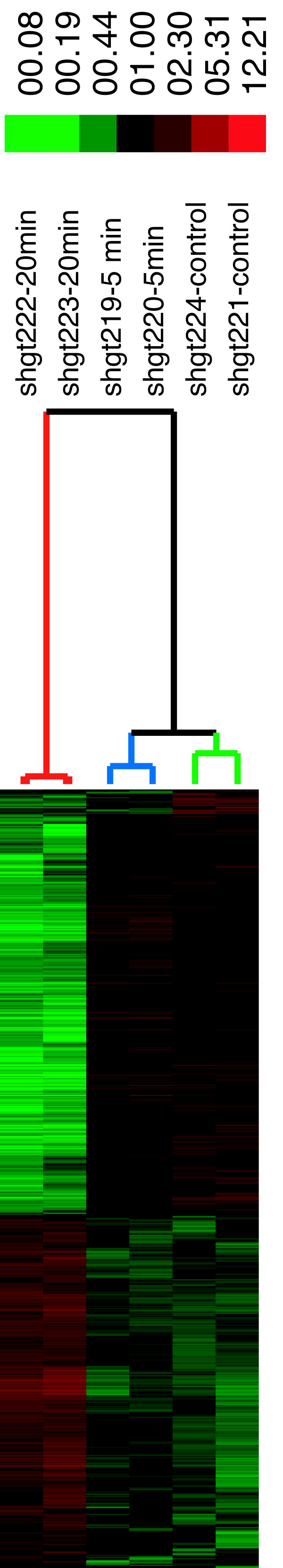
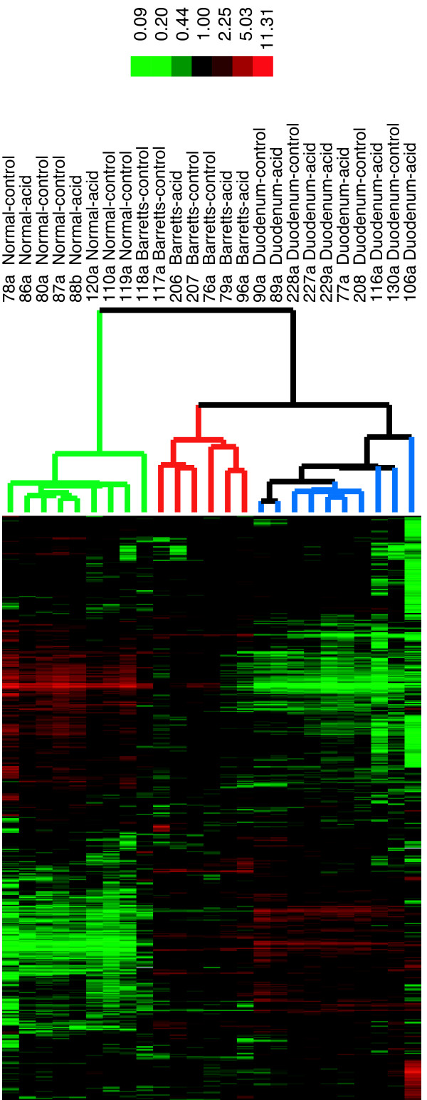
The SEG-1 cell line showed changes in gene expression that was dependent on the length of exposure to pH 3.5. Further analysis using the Gene Ontology, however, showed that representation by genes associated with cell proliferation is not enhanced by acid exposure. The changes in gene expression also did not involve genes known to be differentially expressed in esophageal adenocarcinoma. Similar experiments using short-term primary cultures of Barrett's esophagus also did not result in detectable changes in gene expression with either acid or bile salt exposure.
Short-term exposure of esophageal adenocarcinoma SEG-1 cells or primary cultures of Barrett's esophagus does not result in gene expression changes that are consistent with enhanced cell proliferation. Thus other model systems are needed that may reflect the impact of acid and bile salt exposure on the esophagus in vivo.
食管反流和巴雷特食管是食管腺癌发生的两个主要危险因素。先前的研究表明,将巴雷特相关的腺癌细胞系SEG-1或巴雷特食管组织的原代培养物短暂暴露于酸或胆汁中会导致与细胞增殖一致的变化。在本研究中,我们确定了类似的酸或胆盐暴露是否会导致基因表达变化,从而深入了解恶性转化。
采用先前发表的方法,将巴雷特相关的食管腺癌细胞系和巴雷特食管组织的原代培养物短暂暴露于酸或胆盐脉冲下,然后在pH 7.4的培养基中孵育。然后使用cDNA微阵列对样品进行全基因组基因表达评估。随后的分析评估了处理组和未处理组基因表达的统计学差异。
SEG-1细胞系显示基因表达变化取决于暴露于pH 3.5的时间长度。然而,使用基因本体论的进一步分析表明,酸暴露不会增强与细胞增殖相关基因的表达。基因表达的变化也不涉及已知在食管腺癌中差异表达的基因。使用巴雷特食管短期原代培养物进行的类似实验也未发现酸或胆盐暴露导致基因表达有可检测的变化。
食管腺癌SEG-1细胞或巴雷特食管原代培养物的短期暴露不会导致与细胞增殖增强一致的基因表达变化。因此,需要其他模型系统来反映酸和胆盐暴露对体内食管的影响。