Zhang S, Lin L, Kaur S, Thankachan S, Blanco-Centurion C, Yanagisawa M, Mignot E, Shiromani P J
Stanford University, 701 Welch Road, Room 145, Palo Alto, CA 94304-5742, USA.
Neuroscience. 2007 Aug 10;148(1):34-43. doi: 10.1016/j.neuroscience.2007.05.029. Epub 2007 Jul 6.
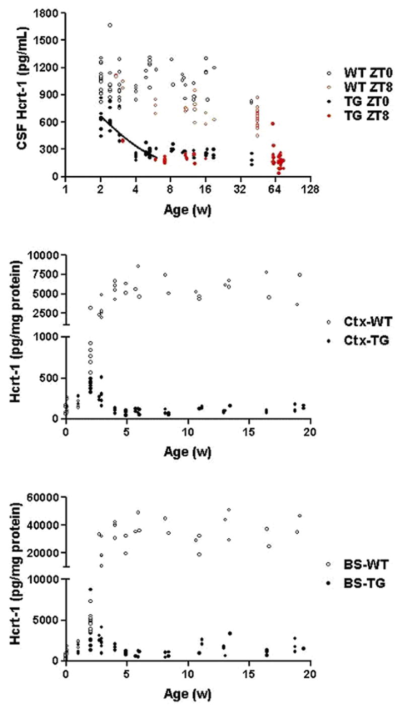
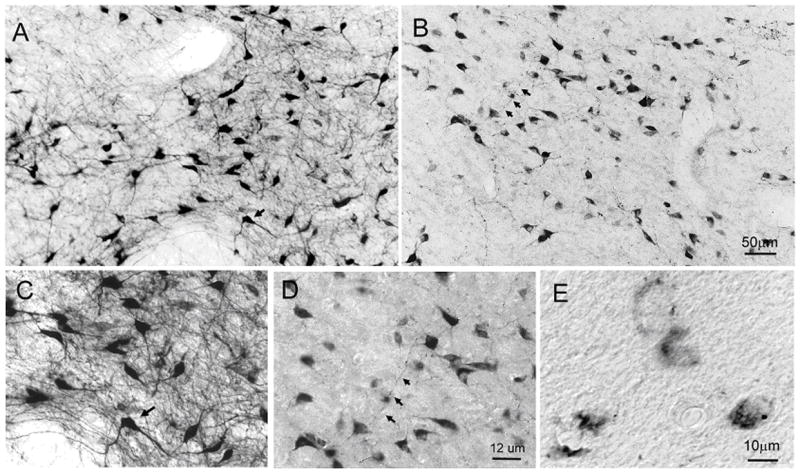
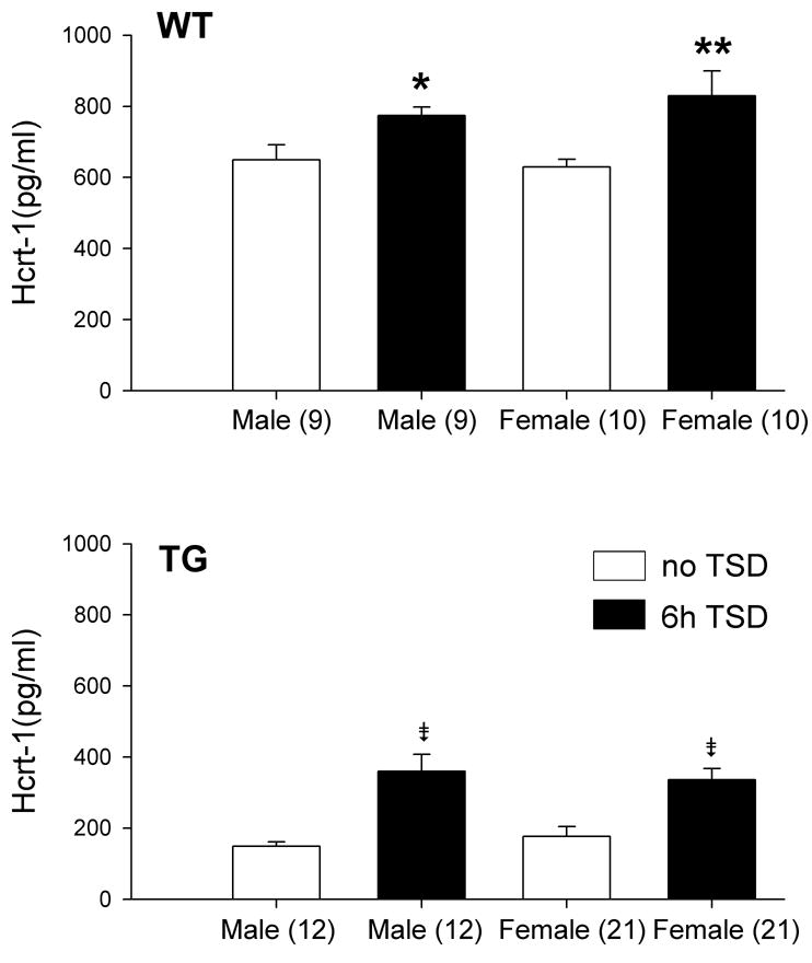
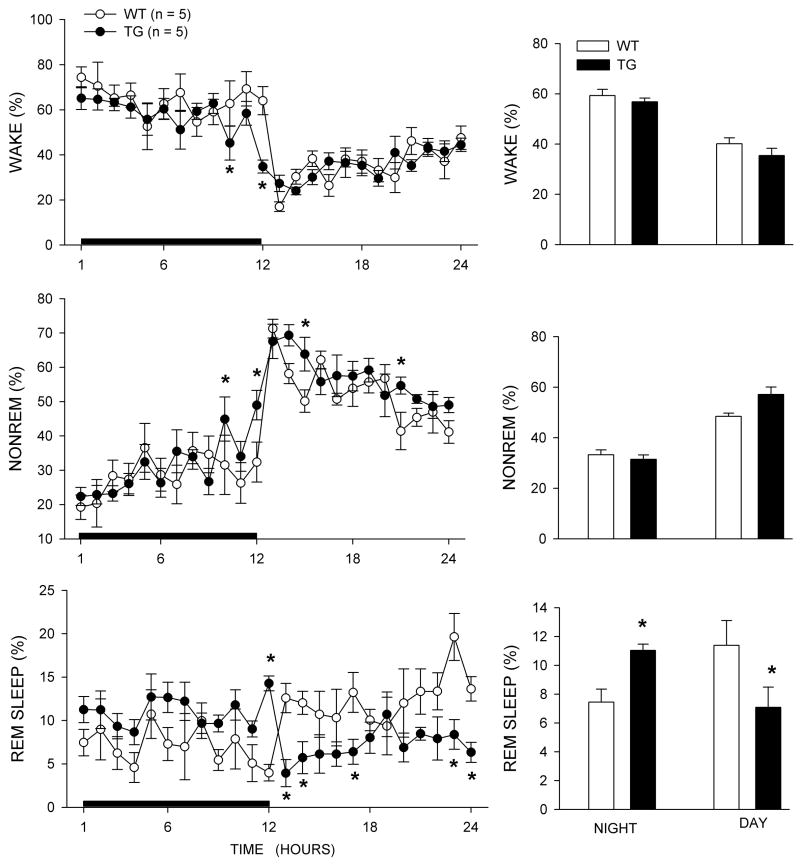
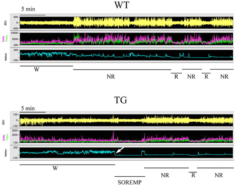
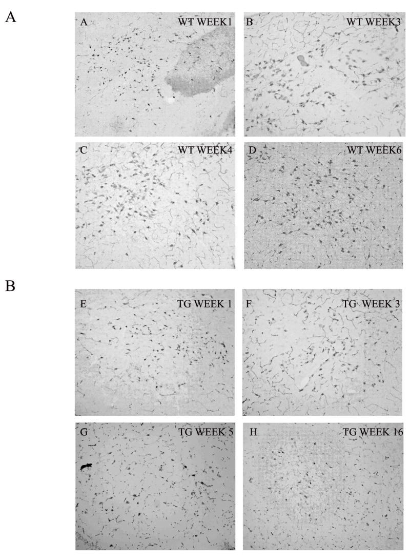
Narcolepsy is linked to a widespread loss of neurons containing the neuropeptide hypocretin (HCRT), also named orexin. A transgenic (TG) rat model has been developed to mimic the neuronal loss found in narcoleptic humans. In these rats, HCRT neurons gradually die as a result of the expression of a poly-glutamine repeat under the control of the HCRT promoter. To better characterize the changes in HCRT-1 levels in response to the gradual HCRT neuronal loss cerebrospinal fluid (CSF) HCRT-1 levels were measured in various age groups (2-82 weeks) of wild-type (WT) and TG Sprague-Dawley rats. TG rats showed a sharp decline in CSF HCRT-1 level at week 4 with levels remaining consistently low (26%+/-9%, mean+/-S.D.) thereafter compared with WT rats. In TG rats, HCRT-1 levels were dramatically lower in target regions such as the cortex and brainstem (100-fold), indicating decreased HCRT-1 levels at terminals. In TG rats, CSF HCRT-1 levels significantly increased in response to 6 h of prolonged waking, indicating that the remaining HCRT neurons can be stimulated to release more neuropeptide. Rapid eye movement (REM) sleep in TG rats (n=5) was consistent with a HCRT deficiency. In TG rats HCRT immunoreactive (HCRT-ir) neurons were present in the lateral hypothalamus (LH), even in old rats (24 months) but some HCRT-ir somata were in various stages of disintegration. The low output of these neurons is consistent with a widespread dysfunction of these neurons, and establishes this model as a tool to investigate the consequences of partial hypocretin deficiency.
发作性睡病与含有神经肽下丘脑分泌素(HCRT,也称为食欲素)的神经元广泛缺失有关。已开发出一种转基因(TG)大鼠模型来模拟发作性睡病患者中发现的神经元缺失。在这些大鼠中,由于在HCRT启动子控制下的多聚谷氨酰胺重复序列的表达,HCRT神经元逐渐死亡。为了更好地表征随着HCRT神经元逐渐缺失,脑脊液(CSF)中HCRT - 1水平的变化,我们在野生型(WT)和TG斯普拉格 - 道利大鼠的不同年龄组(2 - 82周)中测量了CSF中HCRT - 1水平。TG大鼠在第4周时CSF中HCRT - 1水平急剧下降,此后与WT大鼠相比一直保持在较低水平(26%±9%,平均值±标准差)。在TG大鼠中,皮质和脑干等靶区域的HCRT - 1水平显著降低(100倍),表明终末处的HCRT - 1水平降低。在TG大鼠中,延长清醒6小时后,CSF中HCRT - 1水平显著升高,表明剩余的HCRT神经元可被刺激释放更多的神经肽。TG大鼠(n = 5)的快速眼动(REM)睡眠与HCRT缺乏一致。在TG大鼠中,即使是老年大鼠(24个月),下丘脑外侧区(LH)也存在HCRT免疫反应性(HCRT - ir)神经元,但一些HCRT - ir胞体处于不同程度的解体阶段。这些神经元的低输出与这些神经元的广泛功能障碍一致,并将该模型确立为研究部分下丘脑分泌素缺乏后果的工具。