• 文献检索
  • 文档翻译
  • 深度研究
  • 学术资讯
  • Suppr Zotero 插件Zotero 插件
  • 邀请有礼
  • 套餐&价格
  • 历史记录
应用&插件
Suppr Zotero 插件Zotero 插件浏览器插件Mac 客户端Windows 客户端微信小程序
定价
高级版会员购买积分包购买API积分包
服务
文献检索文档翻译深度研究API 文档MCP 服务
关于我们
关于 Suppr公司介绍联系我们用户协议隐私条款
关注我们

Suppr 超能文献

核心技术专利:CN118964589B侵权必究
粤ICP备2023148730 号-1Suppr @ 2026

文献检索

告别复杂PubMed语法,用中文像聊天一样搜索,搜遍4000万医学文献。AI智能推荐,让科研检索更轻松。

立即免费搜索

文件翻译

保留排版,准确专业,支持PDF/Word/PPT等文件格式,支持 12+语言互译。

免费翻译文档

深度研究

AI帮你快速写综述,25分钟生成高质量综述,智能提取关键信息,辅助科研写作。

立即免费体验

相似文献

1
Evaluating computer-aided detection algorithms.评估计算机辅助检测算法。
Med Phys. 2007 Jun;34(6):2024-38. doi: 10.1118/1.2736289.
2
Investigation of reading mode and relative sensitivity as factors that influence reader performance when using computer-aided detection software.研究阅读模式和相对敏感度作为使用计算机辅助检测软件时影响读者表现的因素。
Acad Radiol. 2009 Sep;16(9):1095-107. doi: 10.1016/j.acra.2009.03.024. Epub 2009 Jun 11.
3
A method to test the reproducibility and to improve performance of computer-aided detection schemes for digitized mammograms.一种用于测试数字化乳腺X线摄影计算机辅助检测方案的可重复性并提高其性能的方法。
Med Phys. 2004 Nov;31(11):2964-72. doi: 10.1118/1.1806291.
4
Improving performance of computer-aided detection scheme by combining results from two machine learning classifiers.通过结合两个机器学习分类器的结果来提高计算机辅助检测方案的性能。
Acad Radiol. 2009 Mar;16(3):266-74. doi: 10.1016/j.acra.2008.08.012.
5
On the comparison of FROC curves in mammography CAD systems.乳腺钼靶计算机辅助检测系统中FROC曲线的比较
Med Phys. 2005 Feb;32(2):412-7. doi: 10.1118/1.1844433.
6
GPCALMA: implementation in Italian hospitals of a computer aided detection system for breast lesions by mammography examination.GPCALMA:乳腺钼靶检查中乳腺病变计算机辅助检测系统在意大利医院的应用
Phys Med. 2009 Jun;25(2):58-72. doi: 10.1016/j.ejmp.2008.05.002. Epub 2008 Jul 7.
7
Computer-aided detection; the effect of training databases on detection of subtle breast masses.计算机辅助检测;训练数据库对细微乳腺肿块检测的影响。
Acad Radiol. 2010 Nov;17(11):1401-8. doi: 10.1016/j.acra.2010.06.009. Epub 2010 Jul 22.
8
Computer aided detection of clusters of microcalcifications on full field digital mammograms.全视野数字化乳腺钼靶片上微钙化簇的计算机辅助检测
Med Phys. 2006 Aug;33(8):2975-88. doi: 10.1118/1.2211710.
9
Dual system approach to computer-aided detection of breast masses on mammograms.乳腺钼靶片上乳腺肿块计算机辅助检测的双系统方法。
Med Phys. 2006 Nov;33(11):4157-68. doi: 10.1118/1.2357838.
10
Use of normal tissue context in computer-aided detection of masses in mammograms.利用正常组织上下文辅助计算机检测乳腺 X 线片中的肿块。
IEEE Trans Med Imaging. 2009 Dec;28(12):2033-41. doi: 10.1109/TMI.2009.2028611. Epub 2009 Aug 7.

引用本文的文献

1
Evaluation of computer-aided detection and diagnosis systems.计算机辅助检测和诊断系统的评估。
Med Phys. 2013 Aug;40(8):087001. doi: 10.1118/1.4816310.
2
Application of threshold-bias independent analysis to eye-tracking and FROC data.阈偏独立分析在眼动追踪和 FROC 数据中的应用。
Acad Radiol. 2012 Dec;19(12):1474-83. doi: 10.1016/j.acra.2012.09.002. Epub 2012 Oct 4.
3
Mammographic image denoising and enhancement using the Anscombe transformation, adaptive wiener filtering, and the modulation transfer function.使用安斯科姆变换、自适应维纳滤波和调制传递函数进行乳腺 X 线图像去噪和增强。
J Digit Imaging. 2013 Apr;26(2):183-97. doi: 10.1007/s10278-012-9507-1.
4
Improving the performance of computer-aided detection of subtle breast masses using an adaptive cueing method.利用自适应提示方法提高计算机辅助检测细微乳腺肿块的性能。
Phys Med Biol. 2012 Jan 21;57(2):561-75. doi: 10.1088/0031-9155/57/2/561.
5
Automated detection of multiple sclerosis lesions in serial brain MRI.基于序列脑 MRI 的多发性硬化病变的自动检测。
Neuroradiology. 2012 Aug;54(8):787-807. doi: 10.1007/s00234-011-0992-6. Epub 2011 Dec 20.
6
Improving performance of computer-aided detection of masses by incorporating bilateral mammographic density asymmetry: an assessment.通过纳入双侧乳腺密度不对称来提高计算机辅助检测肿块的性能:评估。
Acad Radiol. 2012 Mar;19(3):303-10. doi: 10.1016/j.acra.2011.10.026. Epub 2011 Dec 14.
7
Computer-assisted detection of infectious lung diseases: a review.计算机辅助检测传染性肺病:综述。
Comput Med Imaging Graph. 2012 Jan;36(1):72-84. doi: 10.1016/j.compmedimag.2011.06.002. Epub 2011 Jul 1.
8
A software framework for building biomedical machine learning classifiers through grid computing resources.一个通过网格计算资源构建生物医学机器学习分类器的软件框架。
J Med Syst. 2012 Aug;36(4):2245-57. doi: 10.1007/s10916-011-9692-3. Epub 2011 Apr 9.
9
Discovering mammography-based machine learning classifiers for breast cancer diagnosis.发现基于乳腺 X 线摄影的机器学习分类器用于乳腺癌诊断。
J Med Syst. 2012 Aug;36(4):2259-69. doi: 10.1007/s10916-011-9693-2. Epub 2011 Apr 9.
10
Computer-aided detection; the effect of training databases on detection of subtle breast masses.计算机辅助检测;训练数据库对细微乳腺肿块检测的影响。
Acad Radiol. 2010 Nov;17(11):1401-8. doi: 10.1016/j.acra.2010.06.009. Epub 2010 Jul 22.

本文引用的文献

1
Receiver operating characteristic analysis: a tool for the quantitative evaluation of observer performance and imaging systems.接收器操作特性分析:一种用于定量评估观察者表现和成像系统的工具。
J Am Coll Radiol. 2006 Jun;3(6):413-22. doi: 10.1016/j.jacr.2006.02.021.
2
Spatial localization accuracy of radiologists in free-response studies: Inferring perceptual FROC curves from mark-rating data.自由反应研究中放射科医生的空间定位准确性:从标记评分数据推断感知FROC曲线。
Acad Radiol. 2007 Jan;14(1):4-18. doi: 10.1016/j.acra.2006.10.015.
3
Multiview-based computer-aided detection scheme for breast masses.基于多视图的乳腺肿块计算机辅助检测方案
Med Phys. 2006 Sep;33(9):3135-43. doi: 10.1118/1.2237476.
4
A completely automated CAD system for mass detection in a large mammographic database.一种用于大型乳腺X线摄影数据库中肿块检测的完全自动化计算机辅助检测系统。
Med Phys. 2006 Aug;33(8):3066-75. doi: 10.1118/1.2214177.
5
Joint two-view information for computerized detection of microcalcifications on mammograms.联合双视图信息用于乳腺钼靶微钙化的计算机检测。
Med Phys. 2006 Jul;33(7):2574-85. doi: 10.1118/1.2208919.
6
ROC curves predicted by a model of visual search.由视觉搜索模型预测的ROC曲线。
Phys Med Biol. 2006 Jul 21;51(14):3463-82. doi: 10.1088/0031-9155/51/14/013. Epub 2006 Jul 6.
7
A search model and figure of merit for observer data acquired according to the free-response paradigm.一种针对根据自由反应范式获取的观察者数据的搜索模型及品质因数。
Phys Med Biol. 2006 Jul 21;51(14):3449-62. doi: 10.1088/0031-9155/51/14/012. Epub 2006 Jul 6.
8
Computer-aided detection in screening CT for pulmonary nodules.用于肺结节筛查的CT中的计算机辅助检测
AJR Am J Roentgenol. 2006 May;186(5):1280-7. doi: 10.2214/AJR.04.1969.
9
Free-response receiver operating characteristic evaluation of lossy JPEG2000 and object-based set partitioning in hierarchical trees compression of digitized mammograms.数字化乳腺X线摄影的有损JPEG2000和基于对象的分层树集分割压缩的自由响应接收器操作特性评估
Radiology. 2005 Nov;237(2):450-7. doi: 10.1148/radiol.2372040996.
10
Computer-aided detection in the United Kingdom National Breast Screening Programme: prospective study.英国国家乳腺筛查计划中的计算机辅助检测:前瞻性研究。
Radiology. 2005 Nov;237(2):444-9. doi: 10.1148/radiol.2372041362.

评估计算机辅助检测算法。

Evaluating computer-aided detection algorithms.

作者信息

Yoon Hong Jun, Zheng Bin, Sahiner Berkman, Chakraborty Dev P

机构信息

Department of Radiology, University of Pittsburgh, Pittsburgh, Pennsylvania 15261, USA.

出版信息

Med Phys. 2007 Jun;34(6):2024-38. doi: 10.1118/1.2736289.

DOI:10.1118/1.2736289
PMID:17654906
原文链接:https://pmc.ncbi.nlm.nih.gov/articles/PMC2041901/
Abstract

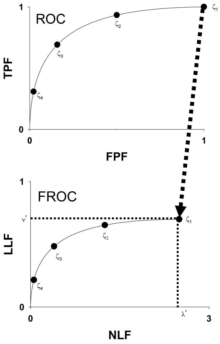
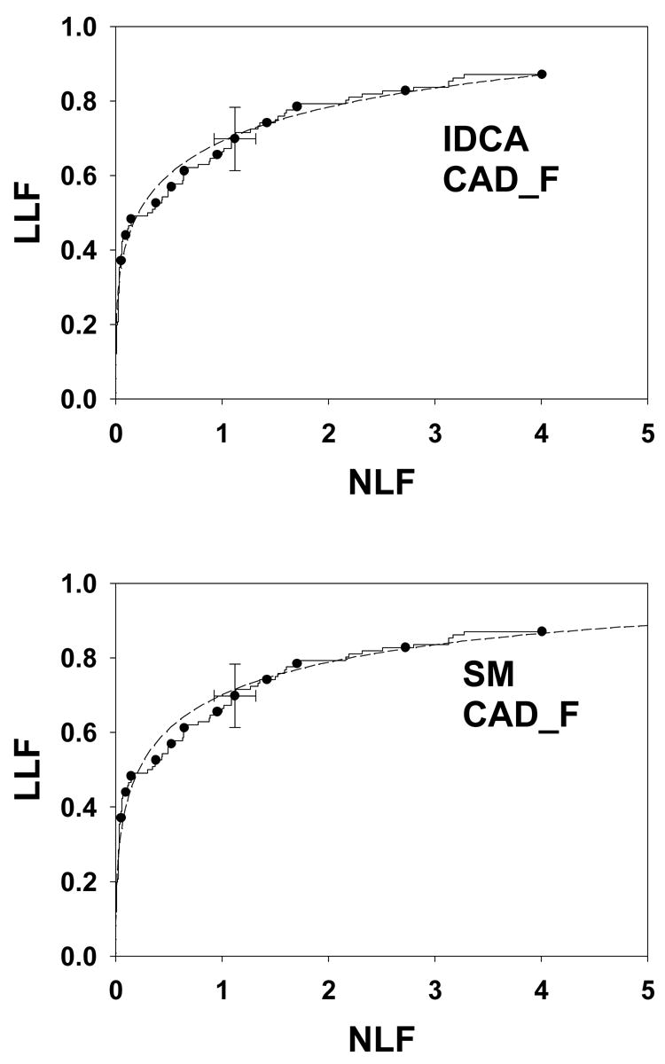
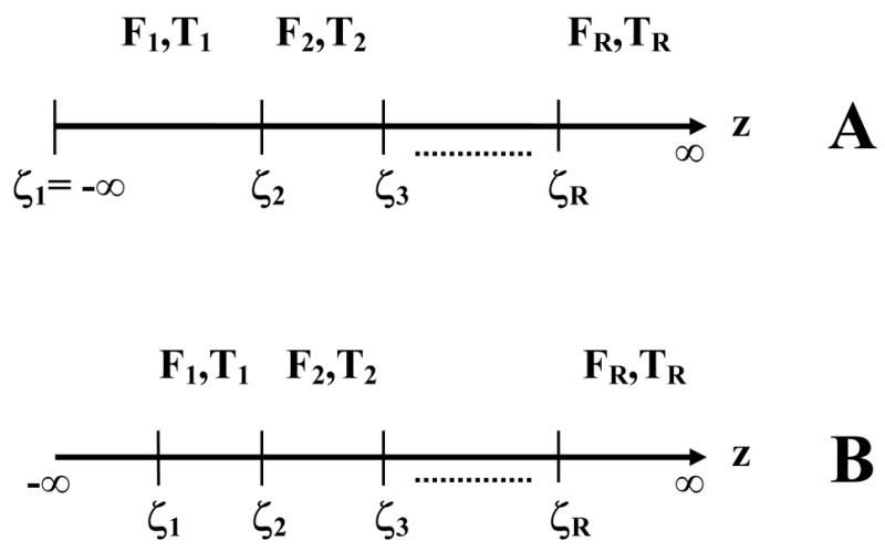
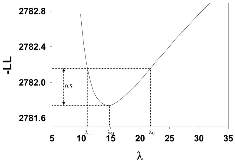
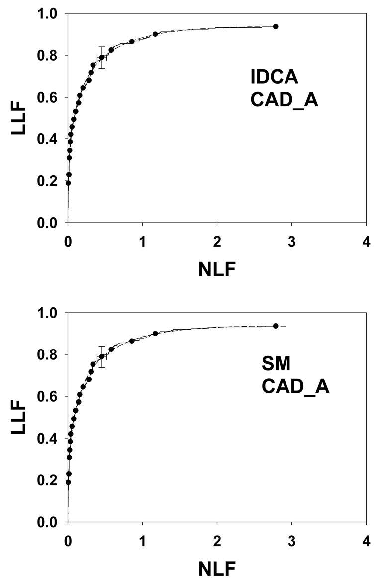
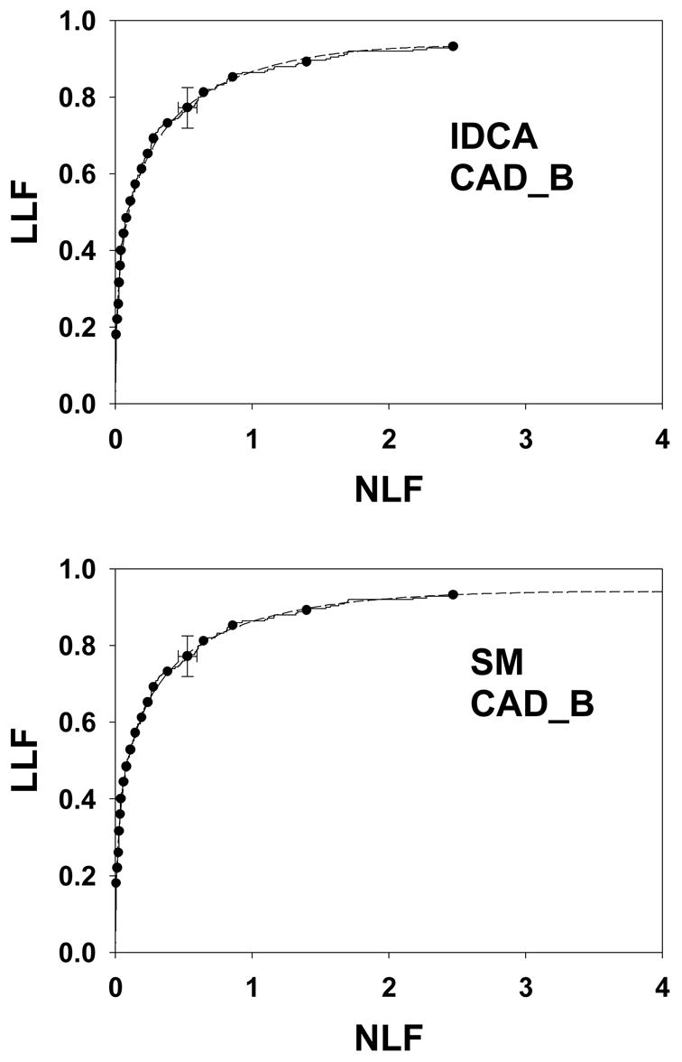
Computer-aided detection (CAD) has been attracting extensive research interest during the last two decades. It is recognized that the full potential of CAD can only be realized by improving the performance and robustness of CAD algorithms and this requires good evaluation methodology that would permit CAD designers to optimize their algorithms. Free-response receiver operating characteristic (FROC) curves are widely used to assess CAD performance, however, evaluation rarely proceeds beyond determination of lesion localization fraction (sensitivity) at an arbitrarily selected value of nonlesion localizations (false marks) per image. This work describes a FROC curve fitting procedure that uses a recent model of visual search that serves as a framework for the free-response task. A maximum likelihood procedure for estimating the parameters of the model from free-response data and fitting CAD generated FROC curves was implemented. Procedures were implemented to estimate two figures of merit and associated statistics such as 95% confidence intervals and goodness of fit. One of the figures of merit does not require the arbitrary specification of an operating point at which to evaluate CAD performance. For comparison a related method termed initial detection and candidate analysis was also implemented that is applicable when all suspicious regions are reported. The two methods were tested on seven mammography CAD data sets and both yielded good to excellent fits. The search model approach has the advantage that it can potentially be applied to radiologist generated free-response data where not all suspicious regions are reported, only the ones that are deemed sufficiently suspicious to warrant clinical follow-up. This work represents the first practical application of the search model to an important evaluation problem in diagnostic radiology. Software based on this work is expected to benefit CAD developers working in diverse areas of medical imaging.

摘要

在过去二十年中,计算机辅助检测(CAD)一直吸引着广泛的研究兴趣。人们认识到,只有通过提高CAD算法的性能和鲁棒性,才能充分发挥CAD的潜力,而这需要良好的评估方法,以便CAD设计人员能够优化其算法。自由响应接收器操作特性(FROC)曲线被广泛用于评估CAD性能,然而,评估很少超出在每张图像任意选定的非病变定位(假标记)值下确定病变定位率(灵敏度)的范围。这项工作描述了一种FROC曲线拟合程序,该程序使用了一种最近的视觉搜索模型,作为自由响应任务的框架。实现了一种从自由响应数据估计模型参数并拟合CAD生成的FROC曲线的最大似然程序。实施了一些程序来估计两个品质因数以及相关统计量,如95%置信区间和拟合优度。其中一个品质因数不需要任意指定评估CAD性能的操作点。为了进行比较,还实施了一种称为初始检测和候选分析的相关方法,该方法适用于报告所有可疑区域的情况。这两种方法在七个乳腺X线摄影CAD数据集上进行了测试,两者都产生了良好到优异的拟合效果。搜索模型方法的优点是,它有可能应用于放射科医生生成的自由响应数据,在这种情况下,并非所有可疑区域都会被报告,只有那些被认为足够可疑以需要临床随访的区域才会被报告。这项工作代表了搜索模型在诊断放射学一个重要评估问题上的首次实际应用。基于这项工作的软件有望使在医学成像不同领域工作的CAD开发人员受益。

https://cdn.ncbi.nlm.nih.gov/pmc/blobs/2c1d/2041901/51d8db6a78a6/nihms24785f6.jpg
https://cdn.ncbi.nlm.nih.gov/pmc/blobs/2c1d/2041901/063be09be357/nihms24785f1.jpg
https://cdn.ncbi.nlm.nih.gov/pmc/blobs/2c1d/2041901/52aa8e4cf0ae/nihms24785f2.jpg
https://cdn.ncbi.nlm.nih.gov/pmc/blobs/2c1d/2041901/5825722fc12d/nihms24785f3.jpg
https://cdn.ncbi.nlm.nih.gov/pmc/blobs/2c1d/2041901/7e9787a54740/nihms24785f4.jpg
https://cdn.ncbi.nlm.nih.gov/pmc/blobs/2c1d/2041901/ab1c7413c5fc/nihms24785f5.jpg
https://cdn.ncbi.nlm.nih.gov/pmc/blobs/2c1d/2041901/51d8db6a78a6/nihms24785f6.jpg