Tuttle Paul V, Rundell Ann E, Webster Thomas J
Weldon School of Biomedical Engineering, Purdue University, West Lafayette, IN 47907, USA.
Int J Nanomedicine. 2006;1(4):497-505. doi: 10.2147/nano.2006.1.4.497.
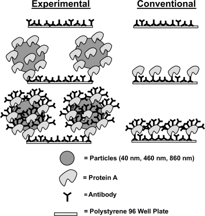
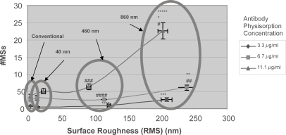
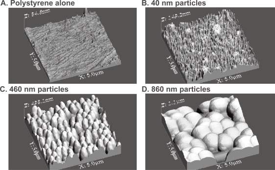
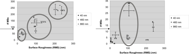
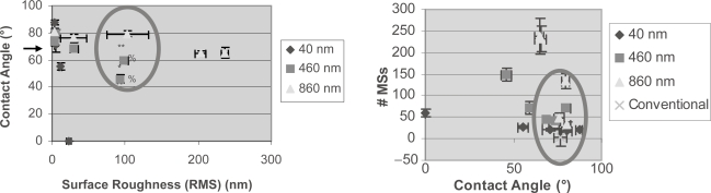
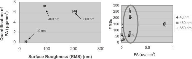
Current research efforts to improve immunoassay-biosensor functionality have centered on detection through the optimal design of microfluidic chambers, electrical circuitry, optical sensing elements, and so on. To date, little attention has been paid to the immunoassay-biosensor membrane surface on which interactions between antibodies and antigens must occur. For this reason, the objective of the present study was to manipulate the nanometer surface roughness of a model immunoassay-biosensor membrane to determine its role on sensitivity and specificity. It was hypothesized that surface roughness characteristics similar to those used by the body's own immune system with B-lymphocyte cell membranes would promote antigen-antibody interactions and minimize non-specific binding. To test this hypothesis, polystyrene 96-well plate surfaces were modified to possess similar topographies as those of B-lymphocyte cell membranes. This was accomplished by immobilizing Protein A conjugated gold particles and Protein A conjugated polystyrene particles ranging in sizes from 40 to 860 nm to the bottom of polystyrene wells. Atomic force microscopy results provided evidence of well-dispersed immunoassay-biosensor surfaces for all particles tested with high degrees of biologically inspired nanometer roughness. Testing the functionality of these immunosurfaces using antigenic fluorescent microspheres showed that specific antigen capture increased with greater nanometer surface roughness while nonspecific antigen capture did not correlate with surface roughness. In this manner, results from this study suggest that large degrees of biologically inspired nanometer surface roughness not only increases the amount of immobilized antibodies onto the immunosurface membrane, but it also enhances the functionality of those antibodies for optimal antigen capture, criteria critical for improving immunoassay-biosensor sensitivity and specificity.
目前,为改善免疫分析生物传感器功能所做的研究工作主要集中在通过微流控腔室、电路、光学传感元件等的优化设计来进行检测。迄今为止,对于免疫分析生物传感器的膜表面(抗体与抗原之间的相互作用必须在此发生)关注甚少。因此,本研究的目的是操控一种模型免疫分析生物传感器膜的纳米表面粗糙度,以确定其对灵敏度和特异性的作用。研究假设是,与人体自身免疫系统中B淋巴细胞细胞膜所具有的表面粗糙度特征相似,将促进抗原 - 抗体相互作用并使非特异性结合最小化。为验证这一假设,对聚苯乙烯96孔板表面进行修饰,使其具有与B淋巴细胞细胞膜相似的形貌。这是通过将尺寸范围为40至860纳米的蛋白A共轭金颗粒和蛋白A共轭聚苯乙烯颗粒固定到聚苯乙烯孔的底部来实现的。原子力显微镜结果为所有测试颗粒提供了免疫分析生物传感器表面分散良好的证据,这些表面具有高度的受生物启发的纳米粗糙度。使用抗原性荧光微球测试这些免疫表面的功能表明,随着纳米表面粗糙度的增加,特异性抗原捕获增加,而非特异性抗原捕获与表面粗糙度无关。通过这种方式,本研究结果表明,高度的受生物启发的纳米表面粗糙度不仅增加了固定在免疫表面膜上的抗体数量,还增强了这些抗体进行最佳抗原捕获的功能,这是提高免疫分析生物传感器灵敏度和特异性的关键标准。