• 文献检索
  • 文档翻译
  • 深度研究
  • 学术资讯
  • Suppr Zotero 插件Zotero 插件
  • 邀请有礼
  • 套餐&价格
  • 历史记录
应用&插件
Suppr Zotero 插件Zotero 插件浏览器插件Mac 客户端Windows 客户端微信小程序
定价
高级版会员购买积分包购买API积分包
服务
文献检索文档翻译深度研究API 文档MCP 服务
关于我们
关于 Suppr公司介绍联系我们用户协议隐私条款
关注我们

Suppr 超能文献

核心技术专利:CN118964589B侵权必究
粤ICP备2023148730 号-1Suppr @ 2026

文献检索

告别复杂PubMed语法,用中文像聊天一样搜索,搜遍4000万医学文献。AI智能推荐,让科研检索更轻松。

立即免费搜索

文件翻译

保留排版,准确专业,支持PDF/Word/PPT等文件格式,支持 12+语言互译。

免费翻译文档

深度研究

AI帮你快速写综述,25分钟生成高质量综述,智能提取关键信息,辅助科研写作。

立即免费体验

心脏瓣膜病:心血管磁共振成像能提供什么信息?

Valvular heart disease: what does cardiovascular MRI add?

作者信息

Masci Pier Giorgio, Dymarkowski Steven, Bogaert Jan

机构信息

Department of Radiology, Gasthuisberg University Hospital, 49 Herestraat, Leuven, 3000, Belgium.

出版信息

Eur Radiol. 2008 Feb;18(2):197-208. doi: 10.1007/s00330-007-0731-x. Epub 2007 Aug 29.

DOI:10.1007/s00330-007-0731-x
PMID:17762934
原文链接:https://pmc.ncbi.nlm.nih.gov/articles/PMC3252017/
Abstract

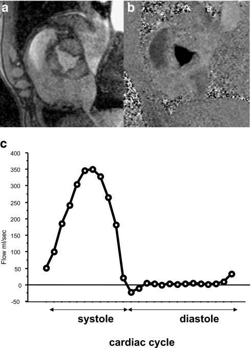
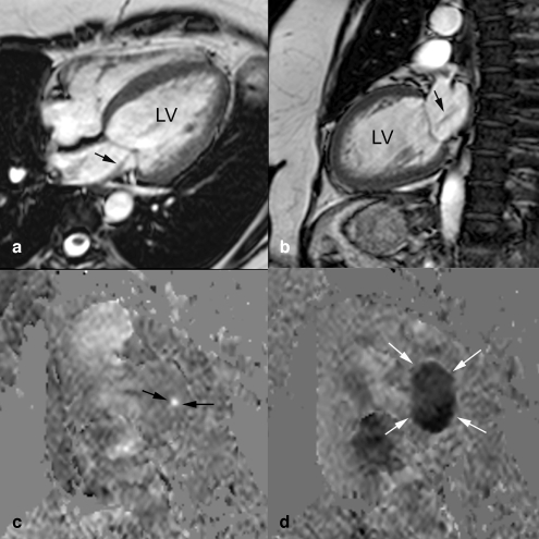
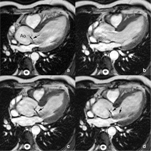
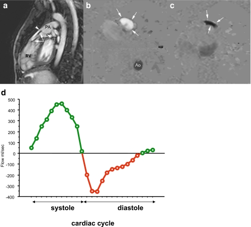
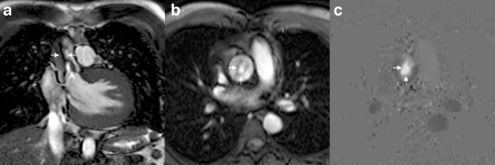
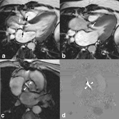
Although ischemic heart disease remains the leading cause of cardiac-related morbidity and mortality in the industrialized countries, a growing number of mainly elderly patients will experience a problem of valvular heart disease (VHD), often requiring surgical intervention at some stage. Doppler-echocardiography is the most popular imaging modality used in the evaluation of this disease entity. It encompasses, however, some non-negligible constraints which may hamper the quality and thus the interpretation of the exam. Cardiac catheterization has been considered for a long time the reference technique in this field, however, this technique is invasive and considered far from optimal. Cardiovascular magnetic resonance imaging (MRI) is already considered an established diagnostic method for studying ventricular dimensions, function and mass. With improvement of MRI soft- and hardware, the assessment of cardiac valve function has also turned out to be fast, accurate and reproducible. This review focuses on the usefulness of MRI in the diagnosis and management of VHD, pointing out its added value in comparison with more conventional diagnostic means.

摘要

尽管在工业化国家,缺血性心脏病仍是心脏相关发病率和死亡率的主要原因,但越来越多的主要是老年患者会出现心脏瓣膜病(VHD)问题,常在某个阶段需要手术干预。多普勒超声心动图是评估这种疾病实体时最常用的成像方式。然而,它存在一些不可忽视的局限性,可能会影响检查质量,进而影响检查结果的解读。长期以来,心导管检查一直被视为该领域的参考技术,然而,这种技术具有侵入性,且远非最佳选择。心血管磁共振成像(MRI)已被认为是研究心室大小、功能和质量的既定诊断方法。随着MRI软硬件的改进,心脏瓣膜功能评估也已变得快速、准确且可重复。本综述重点关注MRI在VHD诊断和管理中的作用,指出其与更传统诊断方法相比的附加价值。

https://cdn.ncbi.nlm.nih.gov/pmc/blobs/d96a/3252017/cb4fe18cb88a/330_2007_731_Fig9_HTML.jpg
https://cdn.ncbi.nlm.nih.gov/pmc/blobs/d96a/3252017/b8afcc34ac6a/330_2007_731_Fig1_HTML.jpg
https://cdn.ncbi.nlm.nih.gov/pmc/blobs/d96a/3252017/4f1cf981883e/330_2007_731_Fig2_HTML.jpg
https://cdn.ncbi.nlm.nih.gov/pmc/blobs/d96a/3252017/9b7f39daf4be/330_2007_731_Fig3_HTML.jpg
https://cdn.ncbi.nlm.nih.gov/pmc/blobs/d96a/3252017/6ff94451687a/330_2007_731_Fig4_HTML.jpg
https://cdn.ncbi.nlm.nih.gov/pmc/blobs/d96a/3252017/f24edf3dd816/330_2007_731_Fig5_HTML.jpg
https://cdn.ncbi.nlm.nih.gov/pmc/blobs/d96a/3252017/6823489ed9a2/330_2007_731_Fig6_HTML.jpg
https://cdn.ncbi.nlm.nih.gov/pmc/blobs/d96a/3252017/a15a79bf06a2/330_2007_731_Fig7_HTML.jpg
https://cdn.ncbi.nlm.nih.gov/pmc/blobs/d96a/3252017/0e58198fe5a4/330_2007_731_Fig8_HTML.jpg
https://cdn.ncbi.nlm.nih.gov/pmc/blobs/d96a/3252017/cb4fe18cb88a/330_2007_731_Fig9_HTML.jpg

相似文献

1
Valvular heart disease: what does cardiovascular MRI add?心脏瓣膜病:心血管磁共振成像能提供什么信息?
Eur Radiol. 2008 Feb;18(2):197-208. doi: 10.1007/s00330-007-0731-x. Epub 2007 Aug 29.
2
Cardiac magnetic resonance imaging in valvular heart disease.心脏磁共振成像在心脏瓣膜病中的应用
Clin Physiol Funct Imaging. 2009 Jul;29(4):229-40. doi: 10.1111/j.1475-097X.2009.00865.x. Epub 2009 Mar 11.
3
Magnetic resonance imaging in valvular heart disease: clinical application and current role for patient management.心脏瓣膜病的磁共振成像:临床应用和患者管理的当前作用。
J Magn Reson Imaging. 2012 Jun;35(6):1241-52. doi: 10.1002/jmri.23544.
4
[Magnetic resonance imaging in the assessment of valvular heart disease].[磁共振成像在心脏瓣膜病评估中的应用]
Rev Esp Cardiol. 2011 Jul;64 Suppl 2:10-8. doi: 10.1016/j.recesp.2011.01.021.
5
[Imaging technique and current status of valvular heart disease using cardiac MRI].[心脏磁共振成像技术在心脏瓣膜病中的应用及现状]
Radiologe. 2013 Oct;53(10):872-9. doi: 10.1007/s00117-012-2468-z.
6
Assessment of valve disease: qualitative and quantitative.瓣膜疾病的评估:定性与定量
Magn Reson Imaging Clin N Am. 2003 Feb;11(1):115-34, vii. doi: 10.1016/s1064-9689(02)00024-7.
7
[Heart valve diseases].[心脏瓣膜疾病]
Radiologe. 1997 May;37(5):341-50. doi: 10.1007/s001170050222.
8
Advanced cardiac MRI techniques for evaluation of left-sided valvular heart disease.用于评估左侧瓣膜性心脏病的先进心脏 MRI 技术。
J Magn Reson Imaging. 2018 Aug;48(2):318-329. doi: 10.1002/jmri.26204.
9
Integrated MR imaging approach to valvular heart disease.瓣膜性心脏病的综合磁共振成像方法。
Cardiol Clin. 1998 May;16(2):277-94. doi: 10.1016/s0733-8651(05)70013-8.
10
The role of cardiac magnetic resonance in valvular heart disease.心脏磁共振在瓣膜性心脏病中的作用。
Methodist Debakey Cardiovasc J. 2013 Jul-Sep;9(3):142-8. doi: 10.14797/mdcj-9-3-142.

引用本文的文献

1
Aortic stenosis assessment from the 3-chamber cine: Ratio of balanced steady-state-free-precession (bSSFP) blood signal between the aorta and left ventricle predicts severity.三腔心电影评估主动脉瓣狭窄:主动脉和左心室之间平衡稳态自由进动(bSSFP)血液信号的比值可预测狭窄严重程度。
J Cardiovasc Magn Reson. 2024 Summer;26(1):100005. doi: 10.1016/j.jocmr.2023.100005. Epub 2024 Jan 9.
2
Cardiovascular Imaging in Pregnancy: Valvulopathy, Hypertrophic Cardiomyopathy, and Aortopathy.孕期心血管成像:瓣膜病、肥厚型心肌病和主动脉病变
Front Cardiovasc Med. 2022 Aug 3;9:834738. doi: 10.3389/fcvm.2022.834738. eCollection 2022.
3

本文引用的文献

1
Quantification of aortic valve stenosis in MRI-comparison of steady-state free precession and fast low-angle shot sequences.MRI 中主动脉瓣狭窄的量化——稳态自由进动和快速低角度激发序列的比较
Eur Radiol. 2007 May;17(5):1284-90. doi: 10.1007/s00330-006-0437-5. Epub 2006 Oct 17.
2
Absolute assessment of aortic valve stenosis by planimetry using cardiovascular magnetic resonance imaging: comparison with transesophageal echocardiography, transthoracic echocardiography, and cardiac catheterisation.使用心血管磁共振成像通过平面测量法对主动脉瓣狭窄进行绝对评估:与经食管超声心动图、经胸超声心动图及心导管检查的比较
Eur J Radiol. 2006 Aug;59(2):276-83. doi: 10.1016/j.ejrad.2006.02.011.
3
Aortic valvular imaging with cardiovascular magnetic resonance: seeking for comprehensiveness.
心血管磁共振成像用于主动脉瓣成像:追求全面性。
Br J Radiol. 2019 Sep;92(1101):20170868. doi: 10.1259/bjr.20170868. Epub 2019 Jul 18.
4
Role of cardiovascular magnetic resonance in acute coronary syndrome.心血管磁共振在急性冠脉综合征中的作用。
Glob Cardiol Sci Pract. 2015 Jul 2;2015(2):24. doi: 10.5339/gcsp.2015.24. eCollection 2015.
5
Quantitative magnetic resonance imaging of pulmonary hypertension: a practical approach to the current state of the art.肺动脉高压的定量磁共振成像:当前技术水平的实用方法
J Thorac Imaging. 2014 Mar;29(2):68-79. doi: 10.1097/RTI.0000000000000079.
6
The role of cardiac magnetic resonance in valvular heart disease.心脏磁共振在瓣膜性心脏病中的作用。
Methodist Debakey Cardiovasc J. 2013 Jul-Sep;9(3):142-8. doi: 10.14797/mdcj-9-3-142.
7
Evaluation of the aortic and mitral valves with cardiac computed tomography and cardiac magnetic resonance imaging.心脏计算机断层扫描和心脏磁共振成像评估主动脉瓣和二尖瓣。
Int J Cardiovasc Imaging. 2012 Dec;28 Suppl 2:109-27. doi: 10.1007/s10554-012-0144-z. Epub 2012 Nov 9.
8
Electrocardiographic QRS duration reflects right ventricular remodeling in patients undergoing corrective surgery for isolated tricuspid regurgitation: a comparative study with cardiac magnetic resonance imaging.心电图 QRS 持续时间反映了孤立性三尖瓣反流矫正手术后患者的右心室重构:与心脏磁共振成像的对比研究。
Clin Cardiol. 2012 Nov;35(11):692-9. doi: 10.1002/clc.22030. Epub 2012 Jun 28.
9
Imaging sequences in cardiovascular magnetic resonance: current role, evolving applications, and technical challenges.心血管磁共振成像序列:当前的作用、不断发展的应用和技术挑战。
Int J Cardiovasc Imaging. 2012 Dec;28(8):2027-47. doi: 10.1007/s10554-012-0038-0. Epub 2012 Mar 25.
10
Planimetric measurement of the regurgitant orifice area using multidetector CT for aortic regurgitation: a comparison with the use of echocardiography.应用多排 CT 进行主动脉瓣反流的瓣口面积平面测量:与超声心动图的比较。
Korean J Radiol. 2010 Mar-Apr;11(2):169-77. doi: 10.3348/kjr.2010.11.2.169. Epub 2010 Feb 22.
Dual-source CT cardiac imaging: initial experience.
双源CT心脏成像:初步经验
Eur Radiol. 2006 Jul;16(7):1409-15. doi: 10.1007/s00330-006-0298-y. Epub 2006 May 13.
4
Severity of mitral and aortic regurgitation as assessed by cardiovascular magnetic resonance: optimizing correlation with Doppler echocardiography.通过心血管磁共振评估二尖瓣和主动脉瓣反流的严重程度:优化与多普勒超声心动图的相关性
J Cardiovasc Magn Reson. 2006;8(3):503-7. doi: 10.1080/10976640600604856.
5
Assessment of magnetic field (4.7 T) induced forces on prosthetic heart valves and annuloplasty rings.评估磁场(4.7特斯拉)对人工心脏瓣膜和瓣环成形环产生的力。
J Magn Reson Imaging. 2005 Aug;22(2):311-7. doi: 10.1002/jmri.20373.
6
Advances in cardiac CT imaging: 64-slice scanner.心脏CT成像进展:64层扫描仪
Int J Cardiovasc Imaging. 2004 Dec;20(6):535-40. doi: 10.1007/s10554-004-7015-1.
7
Magnetic resonance imaging of coronary arteries, the ischemic cascade, and myocardial infarction.冠状动脉、缺血级联反应和心肌梗死的磁共振成像。
Am Heart J. 2005 Feb;149(2):200-8. doi: 10.1016/j.ahj.2004.07.033.
8
Quantitative determinants of the outcome of asymptomatic mitral regurgitation.无症状性二尖瓣反流结局的定量决定因素。
N Engl J Med. 2005 Mar 3;352(9):875-83. doi: 10.1056/NEJMoa041451.
9
Assessment of regional left ventricular function: accuracy and reproducibility of positioning standard short-axis sections in cardiac MR imaging.左心室局部功能评估:心脏磁共振成像中定位标准短轴切面的准确性和可重复性
Radiology. 2005 Apr;235(1):229-36. doi: 10.1148/radiol.2351040249. Epub 2005 Feb 24.
10
Quantification of regurgitant fraction in mitral regurgitation by cardiovascular magnetic resonance: comparison of techniques.心血管磁共振定量二尖瓣反流的反流分数:技术比较
J Heart Valve Dis. 2004 Jul;13(4):600-7.