Loimaala Antti, Groundstroem Kaj, Rinne Marjo, Nenonen Arja, Huhtala Heini, Vuori Ilkka
Clinical Physiology and Nuclear Medicine, Seinäjoki Central Hospital, Seinäjoki, Finland.
Cardiovasc Ultrasound. 2007 Sep 26;5:32. doi: 10.1186/1476-7120-5-32.
Myocardial diastolic tissue velocities are reduced already in newly onset Type 2 diabetes mellitus (T2D). Poor disease control may lead to left ventricular (LV) systolic dysfunction and heart failure. The aim of this study was to assess the effects of exercise training on myocardial diastolic function in T2D patients without ischemic heart disease.
48 men (52.3 +/- 5.6 yrs) with T2D were randomized to supervised training four times a week and standard therapy (E), or standard treatment alone (C) for 12 months. Glycated hemoglobin (HbA1c), oxygen consumption (VO2max), and muscle strength (Sit-up) were measured. Tissue Doppler Imaging (TDI) was used to determine the average maximal mitral annular early (Ea) and late (Aa) diastolic as well as systolic (Sa) velocities, systolic strain (epsilon) and strain rate (epsilon) from the septum, and an estimation of left ventricular end diastolic pressure (E/Ea).
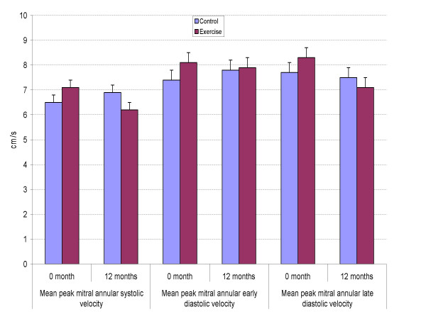
Exercise capacity (VO2max, E 32.0 to 34.7 vs. C 32.6 to 31.5 ml/kg/min, p = .001), muscle strength (E 12.7 to 18.3 times vs. C 14.6 to 14.7 times, p < .001), and HbA1c (E 8.2 to 7.5% vs. C 8.0 to 8.4%, p = .006) improved significantly in the exercise group compared to the controls (ANOVA). Systolic blood pressure decreased in the E group (E 144 to 138 mmHg vs. C 146 to 144 mmHg, p = .04). Contrary to risk factor changes diastolic long axis relaxation did not improve significantly, early diastolic velocity Ea from 8.1 to 7.9 cm/s for the E group vs. C 7.4 to 7.8 cm/s (p = .85, ANOVA). Likewise, after 12 months the mitral annular systolic velocity, systolic strain and strain rate, as well as E/Ea were unchanged.
Exercise training improves endurance and muscle fitness in T2D, resulting in better glycemic control and reduced blood pressure. However, myocardial diastolic tissue velocities did not change significantly. Our data suggest that a much longer exercise intervention may be needed in order to reverse diastolic impairment in diabetics, if at all possible.
在新诊断的2型糖尿病(T2D)患者中,心肌舒张组织速度已经降低。疾病控制不佳可能导致左心室(LV)收缩功能障碍和心力衰竭。本研究的目的是评估运动训练对无缺血性心脏病的T2D患者心肌舒张功能的影响。
48名患有T2D的男性(52.3±5.6岁)被随机分为两组,一组接受每周4次的监督训练和标准治疗(E组),另一组仅接受标准治疗(C组),为期12个月。测量糖化血红蛋白(HbA1c)、耗氧量(VO2max)和肌肉力量(仰卧起坐)。使用组织多普勒成像(TDI)来确定二尖瓣环平均最大舒张早期(Ea)和晚期(Aa)以及收缩期(Sa)速度、室间隔的收缩期应变(ε)和应变率(ε),并估算左心室舒张末期压力(E/Ea)。
与对照组相比,运动组的运动能力(VO2max,E组从32.0提高到34.7,C组从32.6降低到31.5 ml/kg/min,p = 0.001)、肌肉力量(E组从12.7提高到18.3次,C组从14.6提高到14.7次,p < 0.001)和HbA1c(E组从8.2%降低到7.5%,C组从8.0%升高到8.4%,p = 0.006)有显著改善(方差分析)。E组收缩压降低(E组从144 mmHg降至138 mmHg,C组从146 mmHg降至144 mmHg,p = 0.04)。与危险因素变化相反,舒张期长轴松弛没有显著改善,E组舒张早期速度Ea从8.1 cm/s降至7.9 cm/s,C组从7.4 cm/s升至7.8 cm/s(p = 0.85,方差分析)。同样,12个月后二尖瓣环收缩期速度、收缩期应变和应变率以及E/Ea均未改变。
运动训练可改善T2D患者的耐力和肌肉健康,从而更好地控制血糖并降低血压。然而,心肌舒张组织速度没有显著变化。我们的数据表明,如果可能的话,可能需要更长时间的运动干预才能逆转糖尿病患者的舒张功能障碍。