Birmachu Woubalem, Gleason Raymond M, Bulbulian Barbara J, Riter Christie L, Vasilakos John P, Lipson Kenneth E, Nikolsky Yuri
Pharmacology, 3M Pharmaceuticals, St Paul, Minnesota, USA.
BMC Immunol. 2007 Oct 12;8:26. doi: 10.1186/1471-2172-8-26.
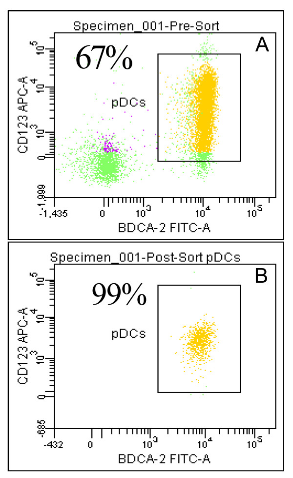
Plasmacytoid Dendritic Cells (pDC) comprise approximately 0.2 to 0.8% of the blood mononuclear cells and are the primary type 1 interferon (IFN), producing cells, secreting high levels of IFN in response to viral infections. Plasmacytoid dendritic cells express predominantly TLRs 7 & 9, making them responsive to ssRNA and CpG DNA. The objective of this study was to evaluate the molecular and cellular processes altered upon stimulation of pDC with synthetic TLR 7 and TLR 7/8 agonists. To this end, we evaluated changes in global gene expression upon stimulation of 99.9% pure human pDC with the TLR7 selective agonists 3M-852A, and the TLR7/8 agonist 3M-011.
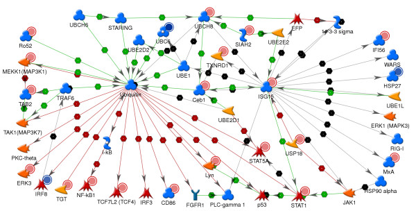
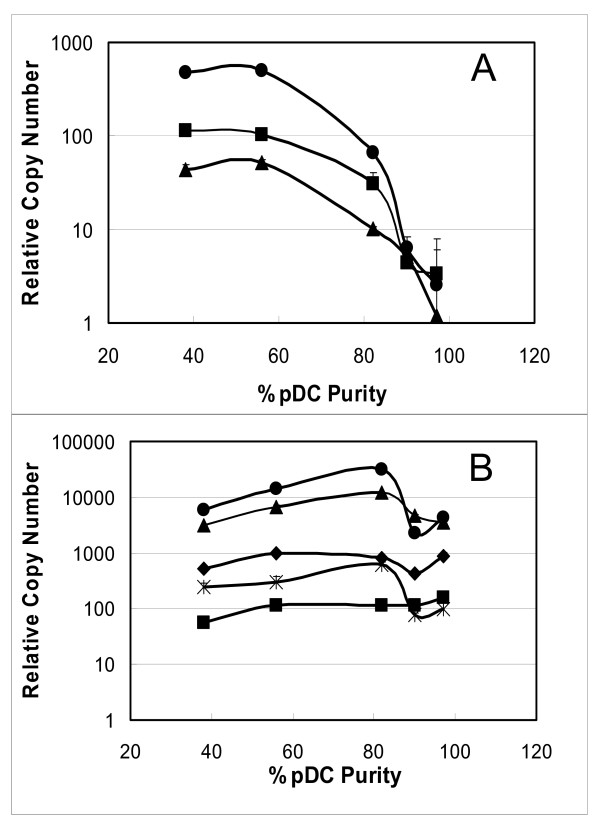
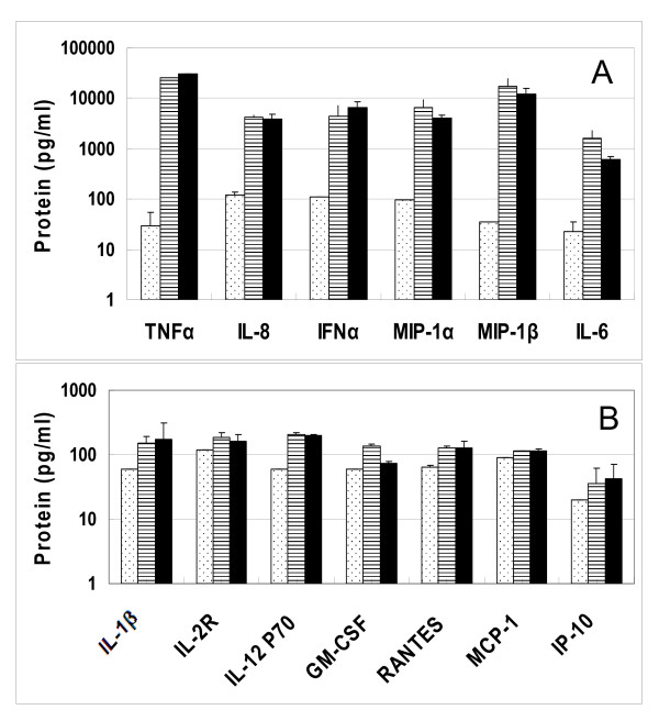
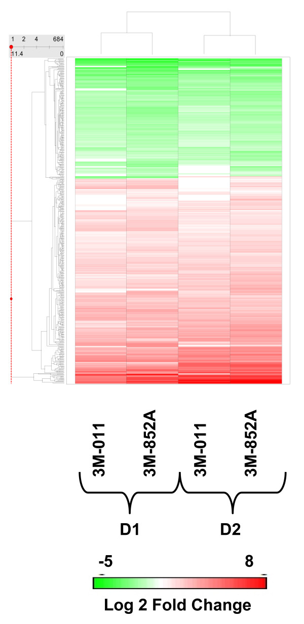
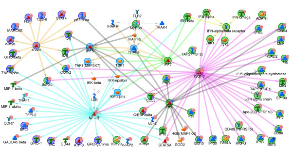
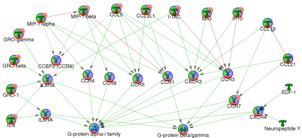
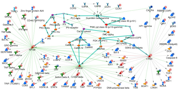
Global gene expression was evaluated using the Affymetrix U133A GeneChip(R) and selected genes were confirmed using real time TaqMan(R) RTPCR. The gene expression profiles of the two agonists were similar indicating that changes in gene expression were solely due to stimulation through TLR7. Type 1 interferons were among the highest induced genes and included IFNB and multiple IFNalpha subtypes, IFNalpha2, alpha5, alpha6, alpha8, alpha1/13, alpha10, alpha14, alpha16, alpha17, alpha21. A large number of chemokines and co-stimulatory molecules as well as the chemokine receptor CCR7 were increased in expression indicating maturation and change in the migratory ability of pDC. Induction of an antiviral state was shown by the expression of several IFN-inducible genes with known anti-viral activity. Further analysis of the data using the pathway analysis tool MetaCore gave insight into molecular and cellular processes impacted. The analysis revealed transcription networks that show increased expression of signaling components in TLR7 and TLR3 pathways, and the cytosolic anti-viral pathway regulated by RIG1 and MDA5, suggestive of optimization of an antiviral state targeted towards RNA viruses. The analysis also revealed increased expression of a network of genes important for protein ISGylation as well as an anti-apoptotic and pro-survival gene expression program.
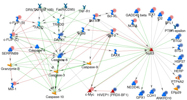
Thus this study demonstrates that as early as 4 hr post stimulation, synthetic TLR7 agonists induce a complex transcription network responsible for activating pDC for innate anti-viral immune responses with optimized responses towards RNA viruses, increased co-stimulatory capacity, and increased survival.
浆细胞样树突状细胞(pDC)约占血液单核细胞的0.2%至0.8%,是主要的1型干扰素(IFN)产生细胞,在病毒感染时分泌高水平的IFN。浆细胞样树突状细胞主要表达Toll样受体(TLR)7和9,使其对单链RNA和CpG DNA有反应。本研究的目的是评估用合成的TLR 7和TLR 7/8激动剂刺激pDC后改变的分子和细胞过程。为此,我们评估了用TLR7选择性激动剂3M-852A和TLR7/8激动剂3M-011刺激99.9%纯的人pDC后全局基因表达的变化。
使用Affymetrix U133A基因芯片评估全局基因表达,并使用实时TaqMan RTPCR确认所选基因。两种激动剂的基因表达谱相似,表明基因表达的变化仅归因于通过TLR7的刺激。1型干扰素是诱导程度最高的基因之一,包括IFNB和多种IFNα亚型,如IFNα2、α5、α6、α8、α1/13、α10、α14、α16、α17、α21。大量趋化因子和共刺激分子以及趋化因子受体CCR7的表达增加,表明pDC的成熟和迁移能力的改变。几种具有已知抗病毒活性的IFN诱导基因的表达显示出抗病毒状态的诱导。使用通路分析工具MetaCore对数据进行的进一步分析深入了解了受影响的分子和细胞过程。分析揭示了转录网络,显示TLR7和TLR3通路中信号成分的表达增加,以及由RIG1和MDA5调节的胞质抗病毒通路,提示针对RNA病毒的抗病毒状态的优化。分析还揭示了对蛋白质ISGylation重要的基因网络以及抗凋亡和促生存基因表达程序的表达增加。
因此,本研究表明,早在刺激后4小时,合成的TLR7激动剂就会诱导一个复杂的转录网络,该网络负责激活pDC以进行先天性抗病毒免疫反应,对RNA病毒的反应得到优化,共刺激能力增加,生存率提高。