Cikos Stefan, Bukovská Alexandra, Koppel Juraj
Institute of Animal Physiology, Slovak Academy of Sciences, Soltésovej 4, 04001 Kosice, Slovakia.
BMC Mol Biol. 2007 Dec 20;8:113. doi: 10.1186/1471-2199-8-113.
Fluorescent data obtained from real-time PCR must be processed by some method of data analysis to obtain the relative quantity of target mRNA. The method chosen for data analysis can strongly influence results of the quantification.
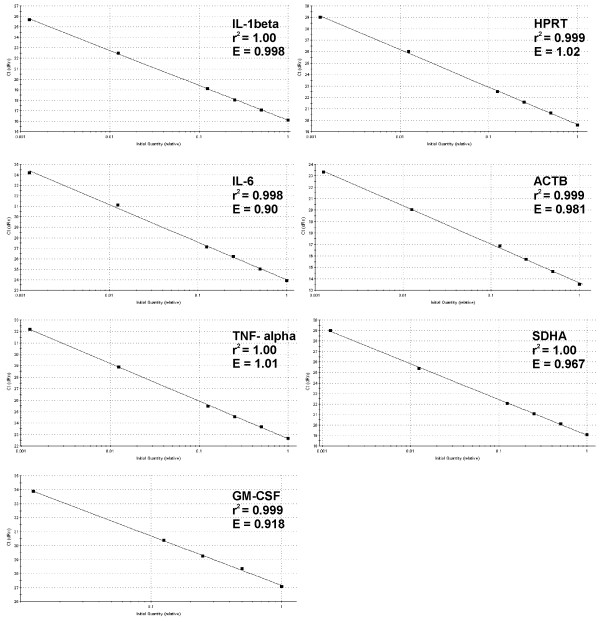
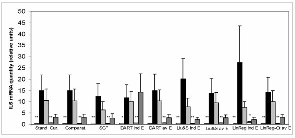
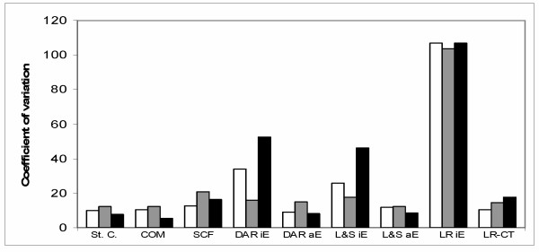
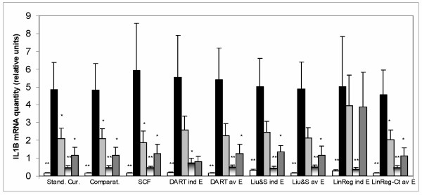
To compare the performance of six techniques which are currently used for analysing fluorescent data in real-time PCR relative quantification, we quantified four cytokine transcripts (IL-1beta, IL-6 TNF-alpha, and GM-CSF) in an in vivo model of colonic inflammation. Accuracy of the methods was tested by quantification on samples with known relative amounts of target mRNAs. Reproducibility of the methods was estimated by the determination of the intra-assay and inter-assay variability. Cytokine expression normalized to the expression of three reference genes (ACTB, HPRT, SDHA) was then determined using the six methods for data analysis. The best results were obtained with the relative standard curve method, comparative Ct method and with DART-PCR, LinRegPCR and Liu & Saint exponential methods when average amplification efficiency was used. The use of individual amplification efficiencies in DART-PCR, LinRegPCR and Liu & Saint exponential methods significantly impaired the results. The sigmoid curve-fitting (SCF) method produced medium performance; the results indicate that the use of appropriate type of fluorescence data and in some instances manual selection of the number of amplification cycles included in the analysis is necessary when the SCF method is applied. We also compared amplification efficiencies (E) and found that although the E values determined by different methods of analysis were not identical, all the methods were capable to identify two genes whose E values significantly differed from other genes.
Our results show that all the tested methods can provide quantitative values reflecting the amounts of measured mRNA in samples, but they differ in their accuracy and reproducibility. Selection of the appropriate method can also depend on the design of a particular experiment. The advantages and disadvantages of the methods in different applications are discussed.
从实时荧光定量PCR获得的荧光数据必须通过某种数据分析方法进行处理,以获得目标mRNA的相对量。选择的数据分析方法会对定量结果产生重大影响。
为比较目前用于实时荧光定量PCR相对定量分析荧光数据的六种技术的性能,我们在结肠炎症的体内模型中对四种细胞因子转录本(IL-1β、IL-6、TNF-α和GM-CSF)进行了定量。通过对已知目标mRNA相对量的样本进行定量来测试方法的准确性。通过测定批内和批间变异性来评估方法的重现性。然后使用六种数据分析方法确定相对于三个参考基因(ACTB、HPRT、SDHA)表达水平进行标准化后的细胞因子表达。当使用平均扩增效率时,相对标准曲线法、比较Ct法以及DART-PCR、LinRegPCR和Liu & Saint指数法获得了最佳结果。在DART-PCR、LinRegPCR和Liu & Saint指数法中使用个体扩增效率会显著损害结果。S形曲线拟合(SCF)法表现中等;结果表明,应用SCF法时,使用适当类型的荧光数据以及在某些情况下手动选择分析中包含的扩增循环数是必要的。我们还比较了扩增效率(E),发现尽管通过不同分析方法确定的E值并不相同,但所有方法都能够识别出两个E值与其他基因有显著差异的基因。
我们的结果表明,所有测试方法都可以提供反映样本中测量mRNA量的定量值,但它们在准确性和重现性方面存在差异。合适方法的选择还可能取决于特定实验的设计。本文讨论了这些方法在不同应用中的优缺点。