Pouliot P, Spahr A, Careau E, Turmel V, Bissonnette E Y
Centre de recherche de l'Hôpital Laval, Institut universitaire de cardiologie et de pneumologie de l'Université Laval, Québec, QC, Canada.
Clin Exp Allergy. 2008 Mar;38(3):529-38. doi: 10.1111/j.1365-2222.2007.02924.x. Epub 2008 Jan 14.
We already demonstrated that adoptive transfer of alveolar macrophages (AMs) from non-allergic rats into AM-depleted allergic rats prevents airway hyperresponsiveness (AHR). We also showed that AMs from non-sensitized, but not from sensitized, allergy-prone rats can prevent AHR following allergen challenge in sensitized allergic animals, establishing the importance of rat immunological status on the modulation of AM functions and suggesting that an allergic lung environment alters AM functions.
We investigated how the activation of allergic AMs can be modulated to reinstitute them with their capacity to reduce AHR.
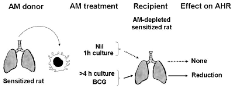
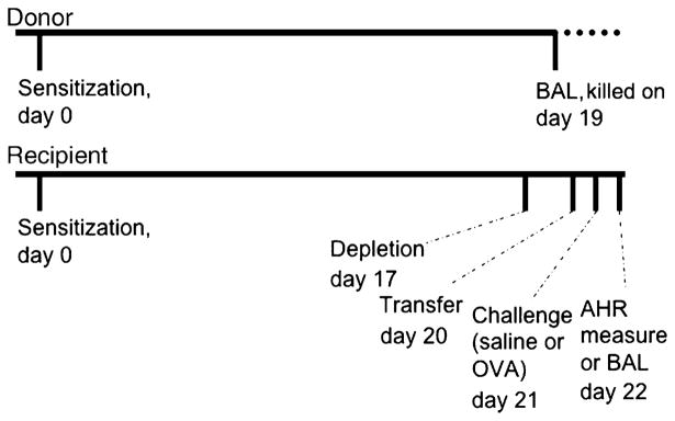
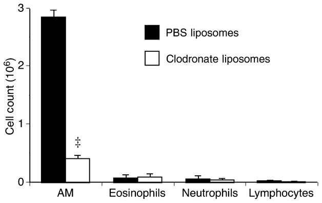
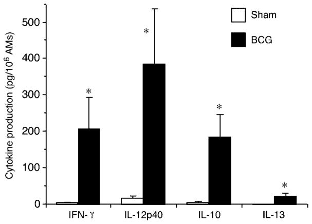
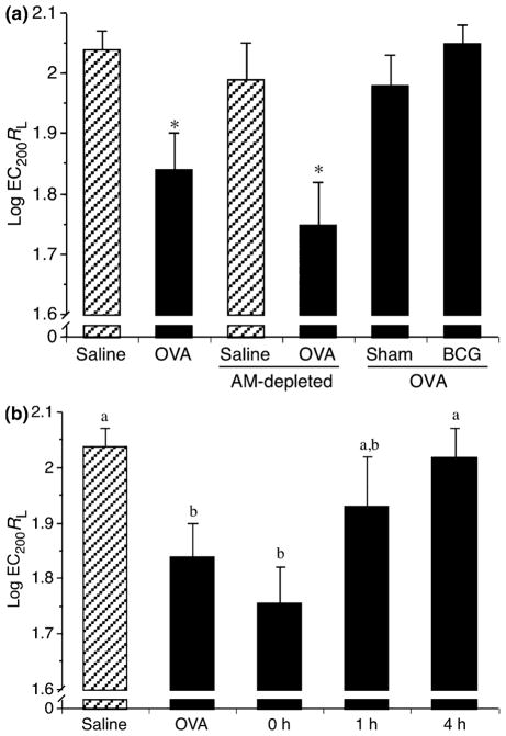
AMs from sensitized Brown Norway rats were cultured ex vivo for up to 18 h in culture media to deprogram them from the influence of the allergic lung before being reintroduced into the lung of AM-depleted sensitized recipient. AHR and cytokines in bronchoalveolar lavage (BAL) were measured following allergen challenge. AMs stimulated ex vivo with Bacillus Calmette-Guerin (BCG) were used as positive controls as BCG induces a T-helper type 1 activation in AMs.
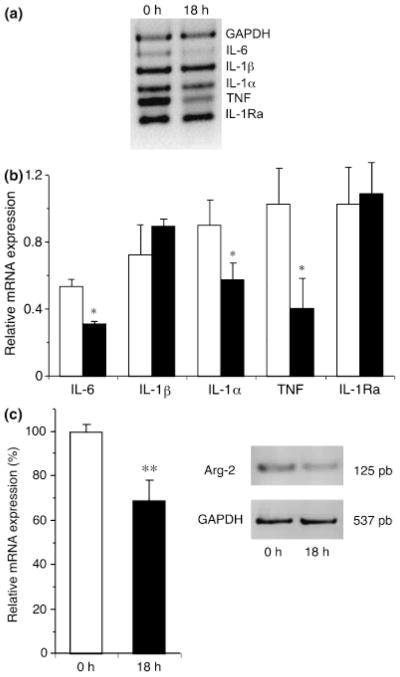
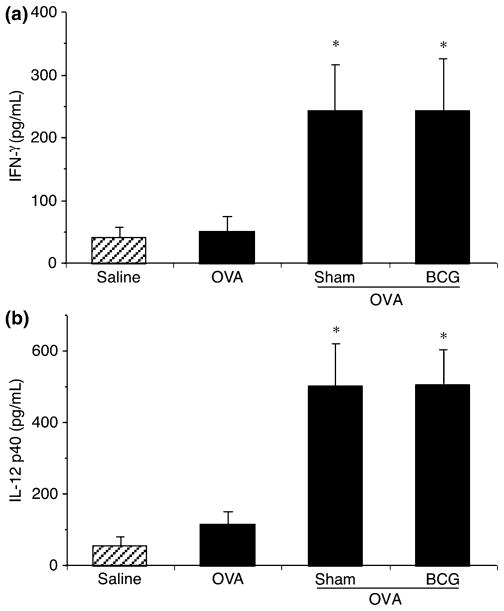
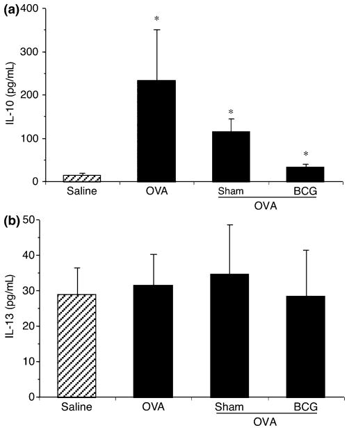
AMs ex vivo cultured for 4-18 h reduced AHR to normal level. Interestingly, pro-allergic functions of AMs were dampened by 18 h culture and they reduced AHR even after spending 48 h in an allergic lung microenvironment. Furthermore, transfer of cultured AMs caused an increase in the levels of IFN-gamma and IL-12 in BAL when compared with their ovalbumin control. After 18 h of ex vivo culture, AMs expressed reduced levels of TNF, IL-1alpha, IL-6, and Arginase-2 mRNAs compared with freshly isolated AMs, suggesting that ex vivo culture exempted AMs from lung stimuli that affected their functions.
There is a significant crosstalk between lung microenvironment and AMs, affecting their functions. It is also the first report showing that sensitized AMs can be modulated ex vivo to reduce lung pro-allergic environment, opening the way to therapies targetting AMs.
我们已经证明,将非过敏性大鼠的肺泡巨噬细胞(AMs)过继转移到AMs耗竭的过敏性大鼠体内可预防气道高反应性(AHR)。我们还表明,来自未致敏但非易致敏的过敏性大鼠的AMs可预防致敏的过敏性动物在过敏原激发后的AHR,这确立了大鼠免疫状态对AMs功能调节的重要性,并表明过敏性肺环境会改变AMs功能。
我们研究了如何调节过敏性AMs的激活,以恢复其降低AHR的能力。
将致敏的棕色挪威大鼠的AMs在培养基中离体培养长达18小时,使其摆脱过敏性肺的影响,然后再重新引入到AMs耗竭的致敏受体的肺中。在过敏原激发后测量支气管肺泡灌洗(BAL)中的AHR和细胞因子。用卡介苗(BCG)离体刺激的AMs用作阳性对照,因为BCG可诱导AMs中的1型辅助性T细胞激活。
离体培养4 - 18小时的AMs将AHR降低至正常水平。有趣的是,AMs的促过敏功能在培养18小时后受到抑制,并且即使在过敏性肺微环境中停留48小时后它们仍能降低AHR。此外,与卵清蛋白对照组相比,培养的AMs的转移导致BAL中IFN - γ和IL - 12水平升高。离体培养18小时后,与新鲜分离的AMs相比,AMs表达的TNF、IL - 1α、IL - 6和精氨酸酶 - 2 mRNA水平降低,这表明离体培养使AMs免受影响其功能的肺刺激。
肺微环境与AMs之间存在显著的相互作用,影响它们的功能。这也是第一项表明致敏的AMs可在离体条件下进行调节以减少肺部促过敏环境的报告,为针对AMs的治疗开辟了道路。