Rakotobe Dina, Violot Sébastien, Hong Saw See, Gouet Patrice, Boulanger Pierre
Laboratoire de Virologie & Pathologie Humaine, Université Lyon I & CNRS FRE-3011, Faculté de Médecine Laennec, 7 rue Guillaume Paradin, 69372 Lyon Cedex 08, France.
Virol J. 2008 Feb 27;5:32. doi: 10.1186/1743-422X-5-32.
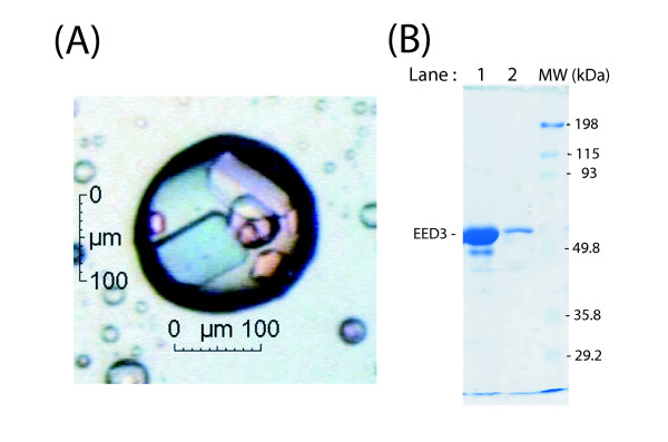
The human EED protein, a member of the superfamily of Polycomb group proteins, is involved in multiple cellular protein complexes. Its C-terminal domain, which is common to the four EED isoforms, contains seven repeats of a canonical WD-40 motif. EED is an interactor of three HIV-1 proteins, matrix (MA), integrase (IN) and Nef. An antiviral activity has been found to be associated with isoforms EED3 and EED4 at the late stage of HIV-1 replication, due to a negative effect on virus assembly and genomic RNA packaging. The aim of the present study was to determine the regions of the EED C-terminal core domain which were accessible and available to protein interactions, using three-dimensional (3D) protein homology modelling with a WD-40 protein of known structure, and epitope mapping of anti-EED antibodies.
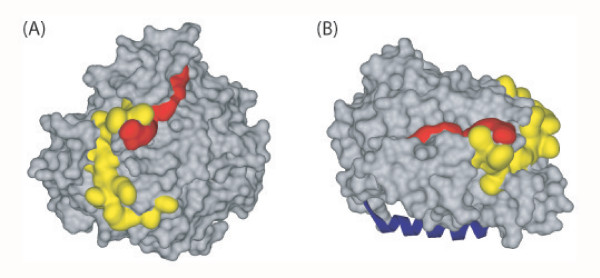
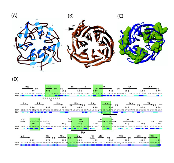
Our data suggested that the C-terminal domain of EED was folded as a seven-bladed beta-propeller protein. During the completion of our work, crystallographic data of EED became available from co-crystals of the EED C-terminal core with the N-terminal domain of its cellular partner EZH2. Our 3D-model was in good congruence with the refined structural model determined from crystallographic data, except for a unique alpha-helix in the fourth beta-blade. More importantly, the position of flexible loops and accessible beta-strands on the beta-propeller was consistent with our mapping of immunogenic epitopes and sites of interaction with HIV-1 MA and IN. Certain immunoreactive regions were found to overlap with the EZH2, MA and IN binding sites, confirming their accessibility and reactivity at the surface of EED. Crystal structure of EED showed that the two discrete regions of interaction with MA and IN did not overlap with each other, nor with the EZH2 binding pocket, but were contiguous, and formed a continuous binding groove running along the lateral face of the beta-propeller.
Identification of antibody-, MA-, IN- and EZH2-binding sites at the surface of the EED isoform 3 provided a global picture of the immunogenic and protein-protein interacting regions in the EED C-terminal domain, organized as a seven-bladed beta-propeller protein. Mapping of the HIV-1 MA and IN binding sites on the 3D-model of EED core predicted that EED-bound MA and IN ligands would be in close vicinity at the surface of the beta-propeller, and that the occurrence of a ternary complex MA-EED-IN would be possible.
人类EED蛋白是多梳蛋白家族的成员,参与多种细胞蛋白复合物。其C末端结构域是四种EED异构体共有的,包含七个典型WD-40基序的重复序列。EED是三种HIV-1蛋白(基质蛋白(MA)、整合酶(IN)和Nef)的相互作用蛋白。在HIV-1复制后期,已发现EED3和EED4异构体具有抗病毒活性,这是由于它们对病毒组装和基因组RNA包装有负面影响。本研究的目的是利用已知结构的WD-40蛋白进行三维(3D)蛋白质同源建模以及抗EED抗体的表位作图,确定EED C末端核心结构域中可用于蛋白质相互作用的区域。
我们的数据表明,EED的C末端结构域折叠成一个七叶β-螺旋桨蛋白。在我们的工作完成期间,EED的晶体学数据可从EED C末端核心与其细胞伴侣EZH2的N末端结构域的共晶体中获得。除了第四个β叶片中有一个独特的α螺旋外,我们的3D模型与根据晶体学数据确定的精细结构模型高度一致。更重要的是,β-螺旋桨上灵活环和可及β链的位置与我们对免疫原性表位以及与HIV-1 MA和IN相互作用位点的作图一致。发现某些免疫反应区域与EZH2、MA和IN结合位点重叠,证实了它们在EED表面的可及性和反应性。EED的晶体结构表明,与MA和IN相互作用的两个离散区域彼此不重叠,也不与EZH2结合口袋重叠,但相邻,并形成一个沿着β-螺旋桨侧面延伸的连续结合槽。
对EED异构体3表面抗体、MA、IN和EZH2结合位点的鉴定,提供了EED C末端结构域中免疫原性和蛋白质-蛋白质相互作用区域的整体情况,该区域组织成一个七叶β-螺旋桨蛋白。在EED核心的3D模型上对HIV-1 MA和IN结合位点的作图预测,与EED结合的MA和IN配体将在β-螺旋桨表面紧密相邻,并且可能形成三元复合物MA-EED-IN。