de Oliveira David Moraes, de Souza Andrade Emanuel Sávio, da Silveira Márcia Maria Fonseca, Camargo Igor Batista
Postgraduation Program of Oral, Maxillofacial Surgery - School of Dentistry of Pernambuco (FOP/UPE), Brazil.
Int J Med Sci. 2008 Feb 8;5(1):36-40. doi: 10.7150/ijms.5.36.
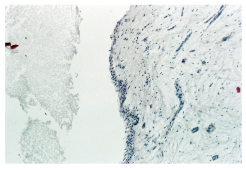
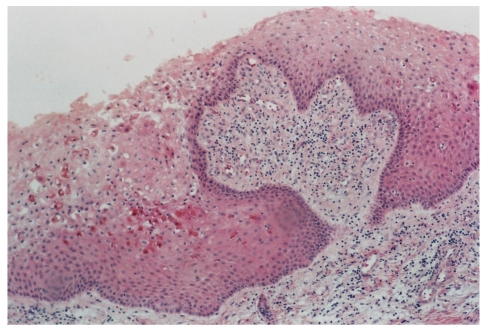
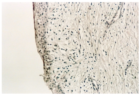
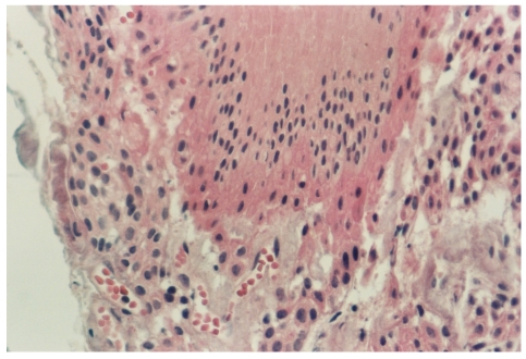
The objective of this study was to determine the correlation of the radiographic and morphological features of the dental follicle of unerupted third molars with incomplete root formation. A cross-sectional study was carried out with 56 patients (105 teeth) aged 13 to 24 years. Panoramic radiography was used to determine the stage of root formation to locate and measure pericoronal radiolucency. The width of the dental follicle ranged from 0.0 to 4.0 mm, the distal face being the one most frequently involved, and stage 7 of root formation showing the highest incidence. An inactive enamel reduced epithelium and inactive epithelium remnant also showed a high incidence. Dense connective tissue showed a high incidence, chronic inflammation was infrequent and calcification was a common finding. There was a significant association between the progression of the rhizogenesis and the transformation of the enamel reduced epithelium into a stratified squamous epithelium. No significant association was found between rhizogenesis and the other morphological findings or between the latter and the width of the pericoronal space. It was concluded that there was no clinically significant correlation between the radiographic and morphological features. Every asymptomatic unerupted third molar should be followed up and the follicular tissue analyzed.
本研究的目的是确定未萌出第三磨牙牙根形成不全时牙囊的影像学特征与形态学特征之间的相关性。对56例年龄在13至24岁的患者(105颗牙齿)进行了横断面研究。采用全景X线摄影确定牙根形成阶段,以定位和测量冠周透射区。牙囊宽度为0.0至4.0mm,远中面受累最为常见,牙根形成第7阶段的发生率最高。无活性釉质减少上皮和无活性上皮残余的发生率也较高。致密结缔组织的发生率较高,慢性炎症少见,钙化常见。牙根形成过程与釉质减少上皮转化为复层鳞状上皮之间存在显著关联。牙根形成与其他形态学表现之间或后者与冠周间隙宽度之间未发现显著关联。结论是影像学特征与形态学特征之间不存在具有临床意义的相关性。每颗无症状的未萌出第三磨牙均应进行随访并分析滤泡组织。