Amrite Aniruddha C, Edelhauser Henry F, Singh Swita R, Kompella Uday B
Department of Pharmaceutical Sciences, University of Nebraska Medical Center, Omaha, NE 68198-5840, USA.
Mol Vis. 2008 Jan 29;14:150-60.
Our previous studies indicated that while 20 nm particles are rapidly cleared from the periocular space of the rat following posterior subconjunctival injection, 200 nm particles persisted for at least two months. To understand faster clearance of 20 nm particles, the purpose of this study was to determine transscleral permeability and in vivo disposition in the presence and absence of circulation. Further, it was the purpose of this study to simulate sustained retinal drug delivery after periocular administration of rapidly cleared and slowly cleared nanoparticles.
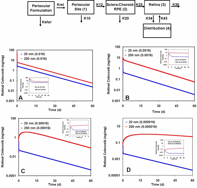
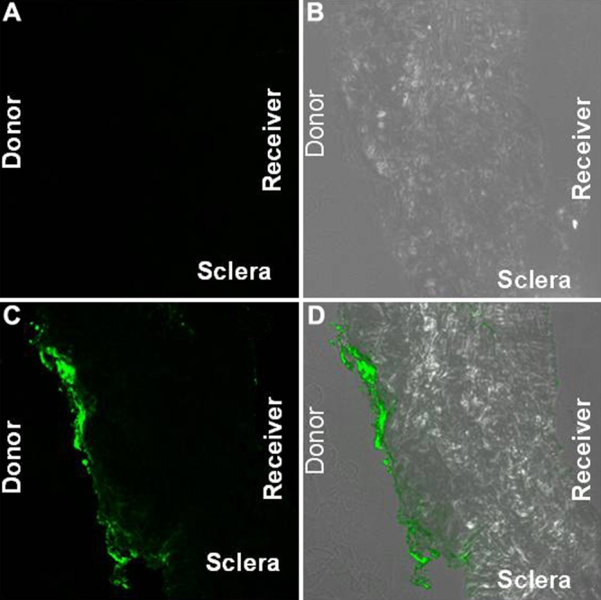
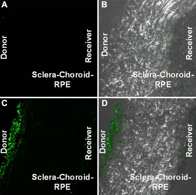
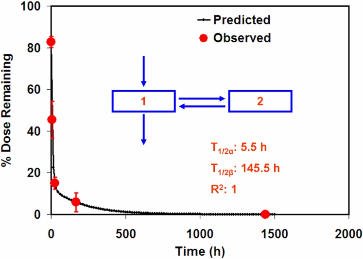
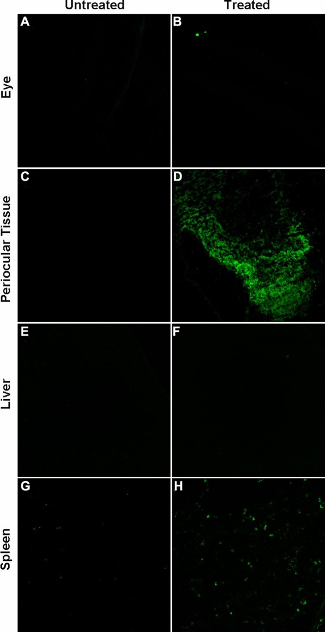
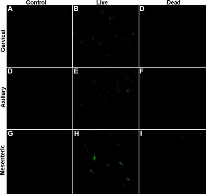
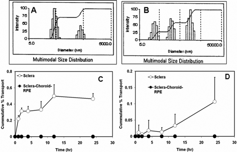
The permeability of 20 and 200 nm particles over 24 h was examined across isolated bovine sclera and sclera-choroid-RPE with or without a surfactant (Tween 20, 0.1% w/v) added to the preparation. The in vivo disposition of nanoparticles was performed using Sprague Dawley rats. The rats, either dead or alive, were administered with 400 microg of the nanoparticles in the periocular space, and the particle disposition in the eye tissues was assessed 6 h later. To evaluate the role of the reticulo-endothelial system and lymphatic circulation, isolated liver, spleen, and cervical, axillary, and mesenteric lymph nodes were analyzed using confocal microscopy. Mathematical simulations with Berkeley Madonna were used to evaluate the effect of nanoparticle size on retinal drug levels following periocular administration. Celecoxib was used as the model drug and the finalized pharmacokinetic model from a previous study was used with some modifications for the simulation.
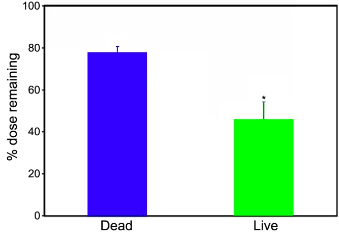
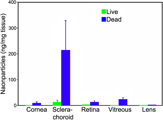
Transport of 20 nm particles across sclera in the presence and absence of the surfactant were 0.1%+/-0.07% and 0.46%+/-0.06%, respectively. These particles did not permeate across the sclera-choroid-RPE in 24 h. There was no quantifiable transport for 200 nm particles across the sclera or the sclera-choroid-RPE. In live animals, the 20 nm particles were undetectable in any of the ocular tissues except in the sclera-choroid following periocular administration; however, in dead animals, the particle concentrations in the sclera-choroid were 19 fold higher than those in live animals, and particles were detectable in the retina as well as vitreous. The retention of 20 nm particles at the site of administration was two fold higher in the dead animals. In live animals, the particles were clearly detectable in the spleen and to a very low extent in the liver as well. The particles were also detected in the cervical, axillary, and mesenteric lymph nodes of the live animals. Simulations with two particles (20 nm and 200 nm) with different clearance rates demonstrated that the retinal drug levels were affected by particle clearance. Larger nanoparticles sustained retinal drug delivery better than smaller nanoparticles. With an increase in drug release rate from the particles, these differences diminish.
The 20 nm particles are transported across the sclera to a minor degree; however, there is no significant transport across the sclera-choroid-RPE. Periocular circulation (blood and lymphatic) plays an important role in the clearance of the 20 nm particles. The higher particle levels in the ocular tissues in the post-mortem studies indicate a dynamic physiologic barrier to the entry of particles into the ocular tissues after periocular administration. The particle size of the delivery system can play an important role in the observed retinal drug levels after periocular administration. Slow release nanoparticles with low clearance by blood and lymphatic circulations are suitable for prolonged transscleral drug delivery to the back of the eye.
我们之前的研究表明,结膜下注射后,20纳米颗粒能迅速从大鼠眼周间隙清除,而200纳米颗粒至少能持续存在两个月。为了解20纳米颗粒更快清除的原因,本研究旨在确定在有循环和无循环情况下的经巩膜通透性及体内分布。此外,本研究的目的是模拟眼周给药后快速清除和缓慢清除的纳米颗粒的持续视网膜药物递送。
在有或无表面活性剂(吐温20,0.1% w/v)添加到制剂的情况下,检测20纳米和200纳米颗粒在24小时内穿过分离的牛巩膜和巩膜-脉络膜-视网膜色素上皮(RPE)的通透性。使用Sprague Dawley大鼠进行纳米颗粒的体内分布研究。对处死或存活的大鼠在眼周间隙给予400微克纳米颗粒,6小时后评估颗粒在眼组织中的分布。为评估网状内皮系统和淋巴循环的作用,使用共聚焦显微镜分析分离的肝脏、脾脏以及颈部、腋窝和肠系膜淋巴结。使用伯克利 Madonna软件进行数学模拟,以评估纳米颗粒大小对眼周给药后视网膜药物水平的影响。使用塞来昔布作为模型药物,并对先前研究中最终确定的药代动力学模型进行一些修改用于模拟。
在有和无表面活性剂的情况下,20纳米颗粒穿过巩膜的转运率分别为0.1%±0.07%和0.46%±0.06%。这些颗粒在24小时内未穿过巩膜-脉络膜-RPE。200纳米颗粒穿过巩膜或巩膜-脉络膜-RPE无可量化的转运。在活体动物中,眼周给药后,除巩膜-脉络膜外,在任何眼组织中均未检测到20纳米颗粒;然而,在处死的动物中,巩膜-脉络膜中的颗粒浓度比活体动物高19倍,且在视网膜和玻璃体中也可检测到颗粒。处死动物中给药部位20纳米颗粒的滞留量高出两倍。在活体动物中,在脾脏中可清楚检测到颗粒,在肝脏中也能检测到但程度非常低。在活体动物的颈部、腋窝和肠系膜淋巴结中也检测到了颗粒。对两种清除率不同的颗粒(20纳米和200纳米)进行模拟表明,视网膜药物水平受颗粒清除的影响。较大的纳米颗粒比较小的纳米颗粒能更好地维持视网膜药物递送。随着颗粒药物释放速率的增加,这些差异减小。
20纳米颗粒仅少量穿过巩膜;然而,穿过巩膜-脉络膜-RPE无明显转运。眼周循环(血液和淋巴)在20纳米颗粒的清除中起重要作用。死后研究中眼组织中较高的颗粒水平表明眼周给药后颗粒进入眼组织存在动态生理屏障。给药系统的颗粒大小对眼周给药后观察到的视网膜药物水平可起重要作用。通过血液和淋巴循环清除率低的缓释纳米颗粒适合经巩膜向眼后部进行长期药物递送。