Danilov Camelia A, Fiskum Gary
Department of Anesthesiology and Program in Neuroscience, University of Maryland School of Medicine, Baltimore, Maryland 21201, USA.
Glia. 2008 May;56(7):801-8. doi: 10.1002/glia.20655.
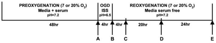
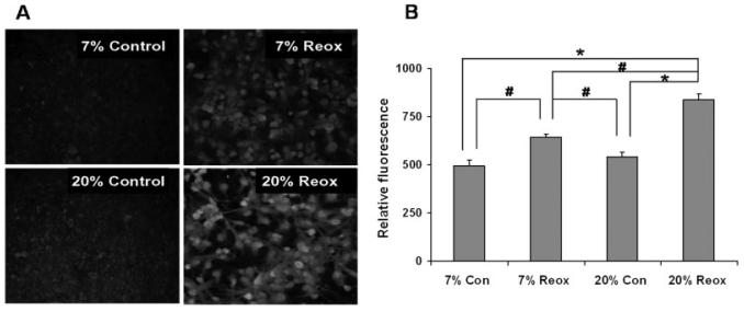
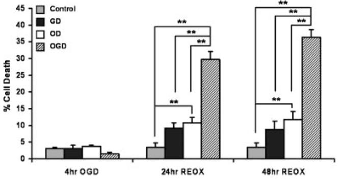
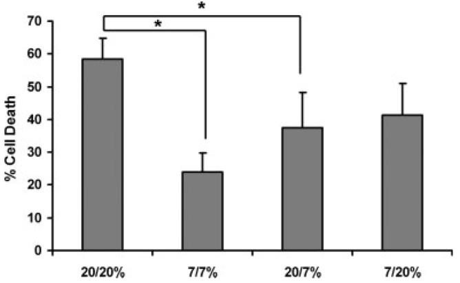
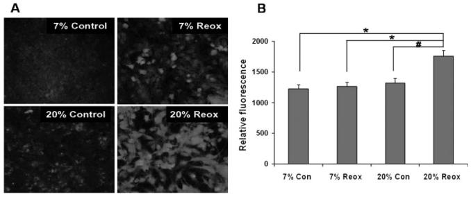
Astrocyte dysfunction and death accompany cerebral ischemia/reperfusion and possibly compromise neuronal survival. Animal studies indicate that neuronal death, neurologic injury, and oxidative molecular modifications are worse in animals exposed to hyperoxic compared to normoxic ventilation during reperfusion after global cerebral ischemia. It is unknown, however, whether ambient O2 affects brain cell survival using in vitro ischemia paradigms where mechanisms of injury to specific cell types can be more thoroughly investigated. This study tested the hypothesis that compared with the supraphysiological level of 20% O2 normally used in cell culture, lower, more physiological O2 levels protect astrocytes from death following oxygen and glucose deprivation. Primary rat cortical astrocytes were cultured under either 7 or 20% O2, exposed to O2, and glucose deprivation for 4 h, and then exposed to normal medium under either 7 or 20% O2. Cell death and 3-nitrotyrosine and 8-hydroxy-2-deoxyguanosine immunoreactivities were assessed at different periods of reoxygenation. Astrocytes exposed to low levels of O2 during reoxygenation undergo less death and exhibit lower levels of protein nitration and nucleic acid oxidation when compared with those under high levels of O2 during reoxygenation. These results support the hypothesis that the 20% O2 normally used in cell culture exacerbates astrocyte death and oxidative stress in an in vitro ischemia/reperfusion model compared to levels that more closely approximate those that exist in vivo.
星形胶质细胞功能障碍和死亡伴随着脑缺血/再灌注,可能会危及神经元的存活。动物研究表明,与全脑缺血再灌注期间常氧通气的动物相比,高氧通气的动物神经元死亡、神经损伤和氧化分子修饰更严重。然而,在体外缺血模型中,环境氧是否会影响脑细胞存活尚不清楚,在该模型中可以更深入地研究特定细胞类型的损伤机制。本研究检验了以下假设:与细胞培养中通常使用的20%超生理水平的氧气相比,更低、更接近生理水平的氧气能保护星形胶质细胞在氧糖剥夺后免于死亡。将原代大鼠皮质星形胶质细胞在7%或20%氧气条件下培养,进行氧糖剥夺4小时,然后在7%或20%氧气条件下置于正常培养基中。在复氧的不同时期评估细胞死亡情况以及3-硝基酪氨酸和8-羟基-2-脱氧鸟苷的免疫反应性。与复氧期间处于高氧水平的星形胶质细胞相比,复氧期间处于低氧水平的星形胶质细胞死亡更少,蛋白质硝化和核酸氧化水平更低。这些结果支持以下假设:与更接近体内存在水平的氧气相比,细胞培养中通常使用的20%氧气在体外缺血/再灌注模型中会加剧星形胶质细胞死亡和氧化应激。