Nordgaard Curtis L, Karunadharma Pabalu P, Feng Xiao, Olsen Timothy W, Ferrington Deborah A
Department of Ophthalmology, University of Minnesota, Minneapolis, Minnesota, USA.
Invest Ophthalmol Vis Sci. 2008 Jul;49(7):2848-55. doi: 10.1167/iovs.07-1352. Epub 2008 Mar 14.
Age-related macular degeneration (AMD) is the leading cause of vision loss in individuals over the age of 65. Histopathological changes become evident in the retinal pigment epithelium (RPE), a monolayer that provides metabolic support for the overlying photoreceptors, even at the earliest stages of AMD that precede vision loss. In a previous global RPE proteome analysis, changes were identified in the content of several mitochondrial proteins associated with AMD. In this study, the subproteome of mitochondria isolated from human donor RPE graded with the Minnesota Grading System (MGS) was analyzed.
Human donor eye bank eyes were categorized into one of four progressive stages (MGS 1-4) based on the clinical features of AMD. After dissection of the RPE, mitochondrial proteins were isolated and separated by two-dimensional gel electrophoresis based on their charge and mass. Protein spot densities were compared between the four MGS stages. Peptides from spots that changed significantly with MGS stage were extracted and analyzed by using mass spectrometry to identify the protein.
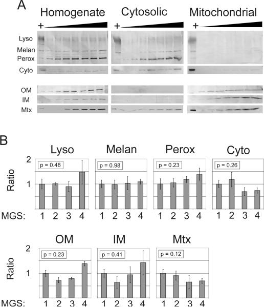
Western blot analyses verified that mitochondria were consistently enriched between MGS stages. The densities of eight spots increased or decreased significantly as a function of MGS stage. These spots were identified as the alpha-, beta-, and delta-ATP synthase subunits, subunit VIb of the cytochrome c oxidase complex, mitofilin, mtHsp70, and the mitochondrial translation factor Tu.
The results are consistent with the hypothesis that mitochondrial dysfunction is associated with AMD and further suggest specific pathophysiological mechanisms involving altered mitochondrial translation, import of nuclear-encoded proteins, and ATP synthase activity.
年龄相关性黄斑变性(AMD)是65岁以上人群视力丧失的主要原因。即使在视力丧失之前的AMD最早阶段,视网膜色素上皮(RPE)(为上方的光感受器提供代谢支持的单层细胞)中的组织病理学变化就已很明显。在先前的一项全球RPE蛋白质组分析中,已确定了几种与AMD相关的线粒体蛋白含量的变化。在本研究中,对从根据明尼苏达分级系统(MGS)分级的人类供体RPE中分离出的线粒体亚蛋白质组进行了分析。
根据AMD的临床特征,将人类供体眼库的眼睛分为四个进展阶段(MGS 1-4)之一。在解剖RPE后,分离线粒体蛋白,并根据其电荷和质量通过二维凝胶电泳进行分离。比较四个MGS阶段之间的蛋白质斑点密度。从随MGS阶段显著变化的斑点中提取肽段,并使用质谱分析以鉴定蛋白质。
蛋白质印迹分析证实,线粒体在MGS阶段之间持续富集。八个斑点的密度随MGS阶段而显著增加或降低。这些斑点被鉴定为α-、β-和δ-ATP合酶亚基、细胞色素c氧化酶复合体的亚基VIb、线粒体融合蛋白、线粒体热休克蛋白70和线粒体翻译因子Tu。
结果与线粒体功能障碍与AMD相关的假设一致,并进一步提示了涉及线粒体翻译改变、核编码蛋白的导入和ATP合酶活性的特定病理生理机制。