Wen Fushi, Celoy Rhodesia M, Nguyen Trang, Zeng Weiqing, Keegstra Kenneth, Immerzeel Peter, Pauly Markus, Hawes Martha C
Division of Plant Pathology and Microbiology, Department of Plant Sciences, Forbes Hall, University of Arizona, Tucson, AZ 85721, USA.
Plant Cell Rep. 2008 Jul;27(7):1125-35. doi: 10.1007/s00299-008-0530-0. Epub 2008 Mar 18.
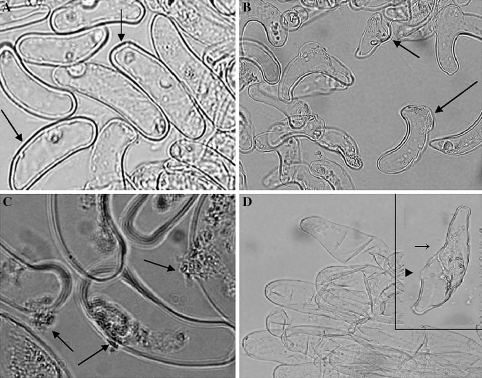
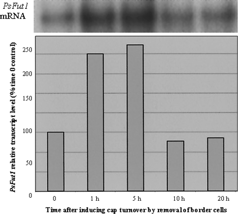
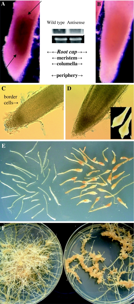
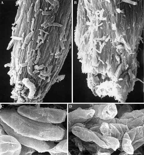
Mitosis and cell wall synthesis in the legume root cap meristem can be induced and synchronized by the nondestructive removal of border cells from the cap periphery. Newly synthesized cells can be examined microscopically as they differentiate progressively during cap development, and ultimately detach as a new population of border cells. This system was used to demonstrate that Pisum sativum L. fucosyl transferase (PsFut1) mRNA expression is strongly expressed in root meristematic tissues, and is induced >2-fold during a 5-h period when mitosis in the root cap meristem is increased. Expression of PsFut1 antisense mRNA in pea hairy roots under the control of the CaMV35S promoter, which exhibits meristem localized expression in pea root caps, resulted in a 50-60% reduction in meristem localized endogenous PsFut1 mRNA expression measured using whole mount in situ hybridization. Changes in gross levels of cell wall fucosylated xyloglucan were not detected, but altered surface localization patterns were detected using whole mount immunolocalization with CCRC-M1, an antibody that recognizes fucosylated xyloglucan. Emerging hairy roots expressing antisense PsFut1 mRNA appeared normal macroscopically but scanning electron microscopy of tissues with altered CCRC-M1 localization patterns revealed wrinkled, collapsed cell surfaces. As individual border cells separated from the cap periphery, cell death occurred in correlation with extrusion of cellular contents through breaks in the wall.
通过从豆科植物根冠外周无损去除边缘细胞,可以诱导并同步根冠分生组织中的有丝分裂和细胞壁合成。新合成的细胞在根冠发育过程中逐渐分化时,可通过显微镜进行观察,最终作为新的边缘细胞群体脱离。该系统用于证明豌豆岩藻糖基转移酶(PsFut1)mRNA在根分生组织中强烈表达,并且在根冠分生组织有丝分裂增加的5小时内诱导表达增加2倍以上。在CaMV35S启动子控制下,豌豆毛状根中PsFut1反义mRNA的表达在豌豆根冠中表现出分生组织定位表达,使用整装原位杂交法测量,导致分生组织定位的内源性PsFut1 mRNA表达降低50 - 60%。未检测到细胞壁岩藻糖基化木葡聚糖总水平的变化,但使用识别岩藻糖基化木葡聚糖的抗体CCRC - M1进行整装免疫定位检测到改变的表面定位模式。表达反义PsFut1 mRNA的新生毛状根在宏观上看起来正常,但对CCRC - M1定位模式改变的组织进行扫描电子显微镜检查发现细胞表面起皱、塌陷。当单个边缘细胞从根冠外周分离时,细胞死亡与细胞内容物通过细胞壁破裂挤出相关。