Strakova Zuzana, Livak Mark, Krezalek Monika, Ihnatovych Ivanna
Department of Obstetrics and Gynecology, University of Illinois at Chicago, Chicago, IL 60612-7313, USA.
Cell Tissue Res. 2008 Jun;332(3):479-88. doi: 10.1007/s00441-008-0604-x. Epub 2008 Apr 10.
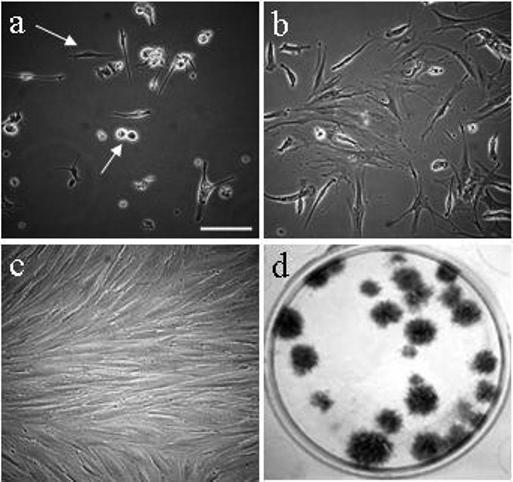
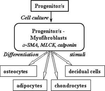
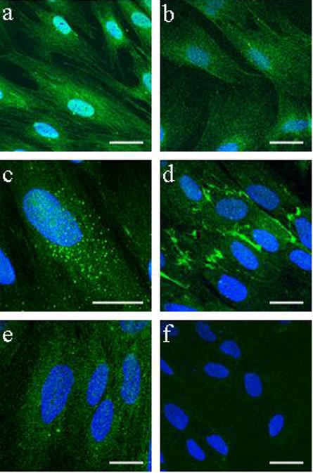
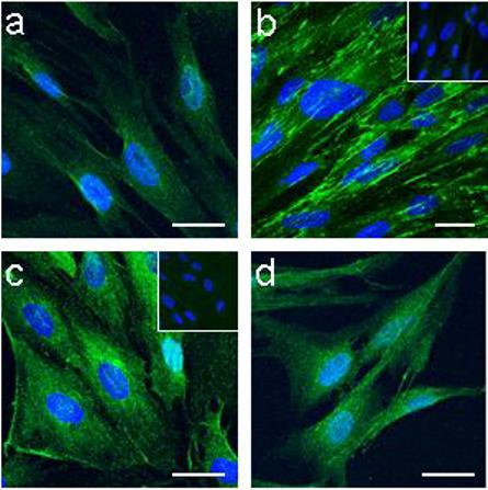
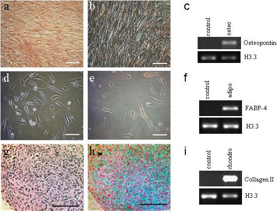
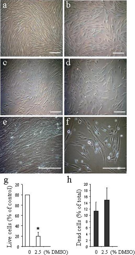
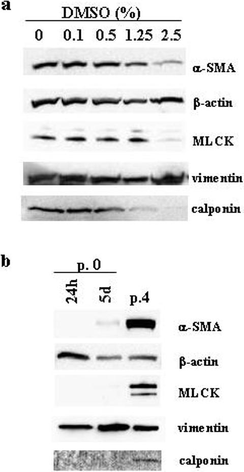
Human uterine fibroblasts (HuF) isolated from the maternal part (decidua parietalis) of a term placenta provide a useful model of in vitro cell differentiation into decidual cells (decidualization, a critical process for successful pregnancy). After isolation, the cells adhere to plastic and have either a small round or spindle-shaped morphology that later changes into a flattened pattern in culture. HuF robustly proliferate in culture until passage 20 and form colonies when plated at low densities. The cells express the mesenchymal cell markers fibronectin, integrin-beta1, ICAM-1 (CD54), and collagen I. Flow cytometry of HuF has detected the presence of CD34, a marker of the hematopoietic stem cell lineage, and an absence of CD10, CD11b/Mac, CD14, CD45, and HLA type II. Furthermore, they also express the pluripotency markers SSEA-1, SSEA-4, Oct-4, Stro-1, and TRA-1-81 as detected by confocal microscopy. Treatment for 14-21 days with differentiation-inducing media leads to the differentiation of HuF into osteoblasts, adipocytes, and chondrocytes. The presence of alpha-smooth muscle actin, calponin, and myosin light-chain kinase in cultured HuF implies their similarity to myofibroblasts. Treatment of the HuF with dimethyl sufoxide causes reversion to the spindle-shaped morphology and a loss of myofibroblast characteristics, suggesting a switch into a less differentiated phenotype. The unique abilities of HuF to exhibit multipotency, even with myofibroblast characteristics, and their ready availability and low maintenance requirements make them an interesting cell model for further exploration as a possible tool for regenerative medicine.
从足月胎盘的母体部分(壁蜕膜)分离出的人子宫成纤维细胞(HuF),为体外细胞分化为蜕膜细胞(蜕膜化,成功妊娠的关键过程)提供了一个有用的模型。分离后,细胞贴附于塑料培养皿,呈小圆形或纺锤形形态,随后在培养过程中变为扁平形态。HuF在培养中强劲增殖直至第20代,低密度接种时形成集落。这些细胞表达间充质细胞标志物纤连蛋白、整合素β1、细胞间黏附分子-1(CD54)和I型胶原。对HuF进行的流式细胞术检测到造血干细胞谱系标志物CD34的存在,且不存在CD10、CD11b/巨噬细胞、CD14、CD45和II类人白细胞抗原。此外,通过共聚焦显微镜检测发现,它们还表达多能性标志物阶段特异性胚胎抗原-1(SSEA-1)、阶段特异性胚胎抗原-4(SSEA-4)、八聚体结合转录因子4(Oct-4)、基质细胞抗原-1(Stro-1)和肿瘤相关抗原-1-81(TRA-1-81)。用诱导分化培养基处理14至21天会导致HuF分化为成骨细胞、脂肪细胞和软骨细胞。培养的HuF中存在α-平滑肌肌动蛋白、钙调蛋白和肌球蛋白轻链激酶,这表明它们与肌成纤维细胞相似。用二甲基亚砜处理HuF会使其恢复为纺锤形形态,并丧失肌成纤维细胞特征,提示转变为分化程度较低的表型。HuF即使具有肌成纤维细胞特征也能表现出多能性的独特能力,以及其易于获取和低维护要求,使其成为作为再生医学可能工具进行进一步探索的有趣细胞模型。