Al-Khayat Hind A, Morris Edward P, Kensler Robert W, Squire John M
Institute of Biomedical Engineering, Imperial College London, Bessemer Building, London SW7 2AZ, UK.
J Struct Biol. 2008 Aug;163(2):117-26. doi: 10.1016/j.jsb.2008.03.011. Epub 2008 Apr 4.
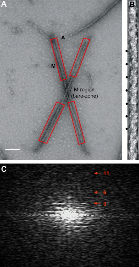
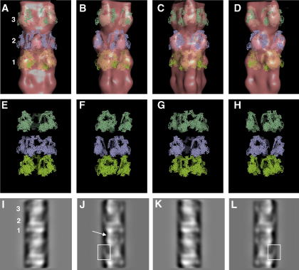
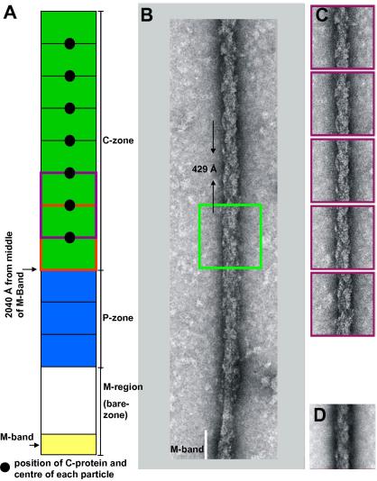
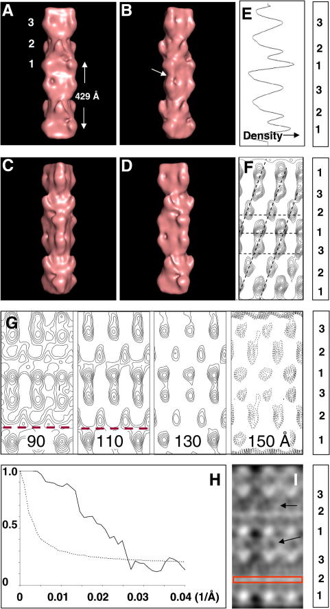
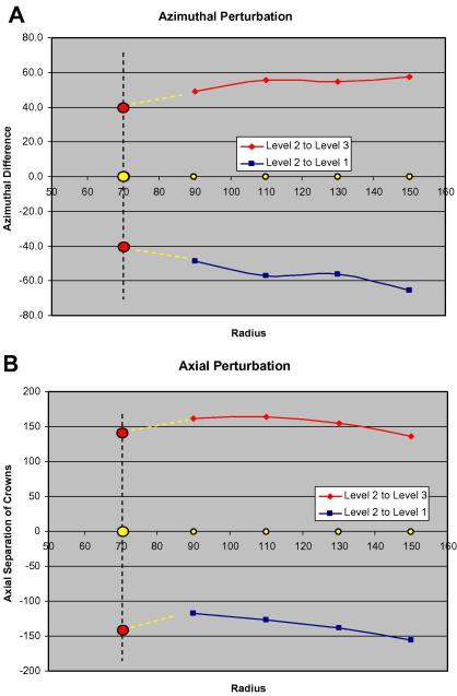
A number of cardiac myopathies (e.g. familial hypertrophic cardiomyopathy and dilated cardiomyopathy) are linked to mutations in cardiac muscle myosin filament proteins, including myosin and myosin binding protein C (MyBP-C). To understand the myopathies it is necessary to know the normal 3D structure of these filaments. We have carried out 3D single particle analysis of electron micrograph images of negatively stained isolated myosin filaments from rabbit cardiac muscle. Single filament images were aligned and divided into segments about 2x430A long, each of which was treated as an independent 'particle'. The resulting 40A resolution 3D reconstruction showed both axial and azimuthal (no radial) myosin head perturbations within the 430A repeat, with successive crown rotations of approximately 60 degrees , 60 degrees and 0 degrees , rather than the regular 40 degrees for an unperturbed helix. However, it is shown that the projecting density peaks appear to start at low radius from origins closer to those expected for an unperturbed helical filament, and that the azimuthal perturbation especially increases with radius. The head arrangements in rabbit cardiac myosin filaments are very similar to those in fish skeletal muscle myosin filaments, suggesting a possible general structural theme for myosin filaments in all vertebrate striated muscles (skeletal and cardiac).
许多心肌病(如家族性肥厚型心肌病和扩张型心肌病)与心肌肌球蛋白丝蛋白的突变有关,包括肌球蛋白和肌球蛋白结合蛋白C(MyBP-C)。为了理解这些疾病,有必要了解这些丝的正常三维结构。我们对来自兔心肌的负染分离肌球蛋白丝的电子显微镜图像进行了三维单颗粒分析。单丝图像被对齐并分成约2×430埃长的片段,每个片段都被视为一个独立的“颗粒”。所得的40埃分辨率三维重建显示,在430埃的重复结构内,肌球蛋白头部在轴向和方位角(无径向)上存在扰动,连续的冠旋转角度约为60度、60度和0度,而不是未受扰动螺旋的规则40度。然而,结果表明,突出的密度峰似乎从离未受扰动螺旋丝预期起始点更近的低半径处开始,并且方位角扰动尤其随半径增加。兔心肌肌球蛋白丝中的头部排列与鱼骨骼肌肌球蛋白丝中的排列非常相似,这表明所有脊椎动物横纹肌(骨骼肌和心肌)中肌球蛋白丝可能存在一个普遍的结构主题。