• 文献检索
  • 文档翻译
  • 深度研究
  • 学术资讯
  • Suppr Zotero 插件Zotero 插件
  • 邀请有礼
  • 套餐&价格
  • 历史记录
应用&插件
Suppr Zotero 插件Zotero 插件浏览器插件Mac 客户端Windows 客户端微信小程序
定价
高级版会员购买积分包购买API积分包
服务
文献检索文档翻译深度研究API 文档MCP 服务
关于我们
关于 Suppr公司介绍联系我们用户协议隐私条款
关注我们

Suppr 超能文献

核心技术专利:CN118964589B侵权必究
粤ICP备2023148730 号-1Suppr @ 2026

文献检索

告别复杂PubMed语法,用中文像聊天一样搜索,搜遍4000万医学文献。AI智能推荐,让科研检索更轻松。

立即免费搜索

文件翻译

保留排版,准确专业,支持PDF/Word/PPT等文件格式,支持 12+语言互译。

免费翻译文档

深度研究

AI帮你快速写综述,25分钟生成高质量综述,智能提取关键信息,辅助科研写作。

立即免费体验

利用 3D 图像分析定义新的牙科表型,以增强对生物过程的区分和深入了解。

Defining new dental phenotypes using 3-D image analysis to enhance discrimination and insights into biological processes.

机构信息

University of Liverpool, Department of Clinical Dental Sciences, Edwards Building, Pembroke Place, Liverpool L69 3GN, UK.

出版信息

Arch Oral Biol. 2009 Dec;54 Suppl 1(Suppl 1):S118-25. doi: 10.1016/j.archoralbio.2008.05.018. Epub 2008 Jul 21.

DOI:10.1016/j.archoralbio.2008.05.018
PMID:18644585
原文链接:https://pmc.ncbi.nlm.nih.gov/articles/PMC2981770/
Abstract

AIMS

In studying aetiological interactions of genetic, epigenetic and environmental factors in normal and abnormal developments of the dentition, methods of measurement have often been limited to maximum mesio-distal and bucco-lingual crown diameters, obtained with hand-held calipers. While this approach has led to many important findings, there are potentially many other informative measurements that can be made to describe dental crown morphology. Advances in digital imaging and computer technology now offer the opportunity to define and measure new dental phenotypes in 3-D that have the potential to provide better anatomical discrimination and clearer insights into the underlying biological processes in dental development. Over recent years, image analysis in 2-D has proved to be a valuable addition to hand-measurement methods but a reliable and rapid 3-D method would increase greatly the morphological information obtainable from natural teeth and dental models. Additional measurements such as crown heights, surface contours, actual surface perimeters and areas, and tooth volumes would maximise our ability to discriminate between samples and to explore more deeply genetic and environmental contributions to observed variation. The research objectives were to investigate the limitations of existing methodologies and to develop and validate new methods for obtaining true 3-D measurements, including curvatures and volumes, in order to enhance discrimination to allow increased differentiation in studies of dental morphology and development. The validity of a new methodology for the 3-D measurement of teeth is compared against an established 2-D system. The intra- and inter-observer reliability of some additional measurements, made possible with a 3-D approach, are also tested.

METHODS AND RESULTS

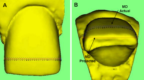
From each of 20 study models, the permanent upper right lateral and upper left central incisors were separated and imaged independently by two operators using 2-D image analysis and a 3-D image analysis system. The mesio-distal (MD), labio-lingual (LL) and inciso-gingival (IG) dimensions were recorded using our 2-D system and the same projected variables were also recorded using a newly developed 3-D system for comparison. Values of Pearson's correlation coefficient between measurements obtained using the two techniques were significant at the 0.01 probability level for variables mesio-distal and incisal-gingival with labio-lingual significant at the 0.05 level for the upper left side only, confirming their comparability. For both 2-D and 3-D systems the intra- and inter-operator reliability was substantial or excellent for variables mesio-distal, labio-lingual, incisal-gingival actual and projected and actual surface area. The reliability was good for inter-operator reliability measurement of the labio-lingual dimension using 3-D.

CONCLUSIONS

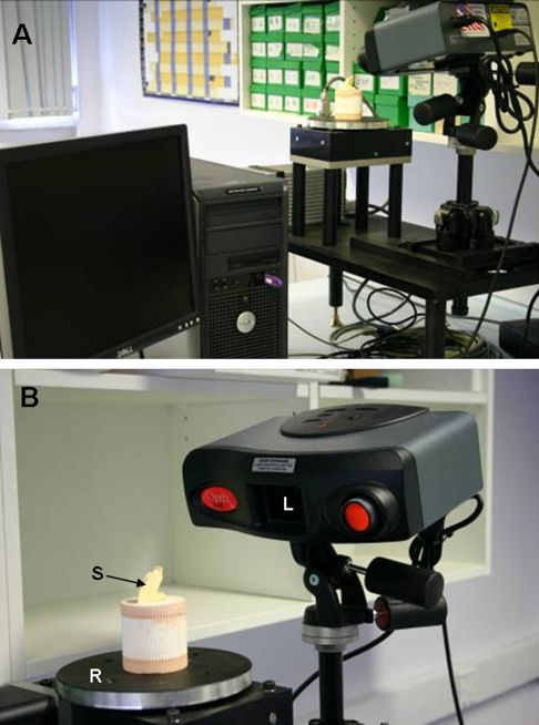
We have developed a new 3-D laser scanning system that enables additional dental phenotypes to be defined. It has been validated against an established 2-D system and shown to provide measurements with excellent reliability, both within and between operators. This new approach provides exciting possibilities for exploring normal and abnormal variations in dental morphology and development applicable to research on genetic and environmental factors.

https://cdn.ncbi.nlm.nih.gov/pmc/blobs/d663/2981770/b77af70ef8ef/gr2.jpg
https://cdn.ncbi.nlm.nih.gov/pmc/blobs/d663/2981770/b575ba674020/gr1.jpg
https://cdn.ncbi.nlm.nih.gov/pmc/blobs/d663/2981770/b77af70ef8ef/gr2.jpg
https://cdn.ncbi.nlm.nih.gov/pmc/blobs/d663/2981770/b575ba674020/gr1.jpg
https://cdn.ncbi.nlm.nih.gov/pmc/blobs/d663/2981770/b77af70ef8ef/gr2.jpg
摘要

目的

在研究遗传、表观遗传和环境因素在牙齿正常和异常发育中的病因学相互作用时,测量方法通常仅限于使用手持卡尺获得的最大近远中向和颊舌向冠直径。虽然这种方法已经带来了许多重要的发现,但实际上还有许多其他有意义的测量方法可以用来描述牙冠形态。数字成像和计算机技术的进步现在为 3D 中的新牙齿表型定义和测量提供了机会,这有可能提供更好的解剖学区分,并更清楚地了解牙齿发育中的潜在生物学过程。近年来,二维图像分析已被证明是对手动测量方法的有益补充,但可靠和快速的 3D 方法将大大增加从天然牙齿和牙齿模型中获得的形态信息量。其他测量方法,如冠高、表面轮廓、实际表面周长和面积以及牙齿体积,将最大限度地提高我们区分样本的能力,并更深入地探索遗传和环境对观察到的变异的贡献。研究目的是研究现有方法的局限性,并开发和验证用于获取真实 3D 测量值的新方法,包括曲率和体积,以增强区分能力,从而允许在牙齿形态和发育研究中进行更大的差异。将一种新的 3D 牙齿测量方法的有效性与一种已建立的 2D 系统进行了比较。还测试了一些额外测量值的 3D 方法的内部和观察者之间的可靠性,这些额外测量值是 3D 方法才能够实现的。

方法和结果

从 20 个研究模型中的每一个中,将右上侧恒侧切牙和左上侧恒中切牙分开,并由两名操作人员分别使用二维图像分析和三维图像分析系统进行独立成像。使用我们的 2D 系统记录近远中(MD)、颊舌向(LL)和切龈向(IG)尺寸,并使用新开发的 3D 系统记录相同的投影变量进行比较。两种技术获得的测量值之间的皮尔逊相关系数值在 0.01 的概率水平上具有统计学意义,MD 和切龈向的变量在 0.05 水平上具有统计学意义,仅在左侧上侧具有统计学意义,证实了它们的可比性。对于 2D 和 3D 系统,对于 MD、LL、IG 的实际和投影以及实际表面面积的变量,内部和观察者之间的可靠性均为中等或良好。对于使用 3D 系统的观察者之间的 LL 尺寸的内部和观察者之间的可靠性测量,可靠性良好。

结论

我们已经开发了一种新的 3D 激光扫描系统,能够定义其他牙齿表型。它已经过与已建立的 2D 系统进行验证,并显示出具有出色的可靠性,无论是在内部还是在操作人员之间。这种新方法为探索牙齿形态和发育的正常和异常变化提供了令人兴奋的可能性,适用于遗传和环境因素的研究。

相似文献

1
Defining new dental phenotypes using 3-D image analysis to enhance discrimination and insights into biological processes.利用 3D 图像分析定义新的牙科表型,以增强对生物过程的区分和深入了解。
Arch Oral Biol. 2009 Dec;54 Suppl 1(Suppl 1):S118-25. doi: 10.1016/j.archoralbio.2008.05.018. Epub 2008 Jul 21.
2
The accuracy of a 3-D laser scanner for crown width measurements.用于测量牙冠宽度的三维激光扫描仪的准确性。
Aust Orthod J. 2009 May;25(1):41-7.
3
Multiple crown size variables of the upper incisors in patients with supernumerary teeth compared with controls.多生牙患者上颌中切牙牙冠各项尺寸变量与对照组的比较。
Arch Oral Biol. 2009 Dec;54 Suppl 1:S71-8. doi: 10.1016/j.archoralbio.2008.10.006. Epub 2008 Dec 19.
4
Quantification of dental plaque on lingual tooth surfaces using image analysis: reliability and validation.使用图像分析对舌侧牙面菌斑进行定量分析:可靠性与验证
J Clin Periodontol. 2004 Jul;31(7):569-73. doi: 10.1111/j.1600-051X.2004.00523.x.
5
[A study on the dental traits and crown sizes of Japanese children].[关于日本儿童牙齿特征及牙冠大小的研究]
Aichi Gakuin Daigaku Shigakkai Shi. 1990 Mar;28(1 Pt 1):159-94.
6
A clinical photogrammetric method to measure dental arch dimensions and mesio-distal tooth size.一种临床摄影测量法,用于测量牙弓尺寸和近远中牙齿尺寸。
Eur J Orthod. 2011 Dec;33(6):721-6. doi: 10.1093/ejo/cjq149. Epub 2011 Jan 31.
7
Fluctuating dental asymmetry of multiple crown variables measured by an image analysis system.通过图像分析系统测量的多个牙冠变量的波动性牙齿不对称性。
Arch Oral Biol. 2005 Feb;50(2):249-53. doi: 10.1016/j.archoralbio.2004.08.013.
8
Mesio-distal tooth angulation in elderly with many remaining teeth observed by 3-D imaging.通过三维成像观察有许多余留牙的老年人的牙近远中倾斜度。
Bull Tokyo Dent Coll. 2010;51(2):57-64. doi: 10.2209/tdcpublication.51.57.
9
Validity, reliability and reproducibility of three methods used to measure tooth widths for bolton analyses.用于Bolton分析的三种测量牙宽度方法的有效性、可靠性和可重复性。
Aust Orthod J. 2009 Nov;25(2):97-103.
10
Tooth dimensions in hypodontia with a known PAX9 mutation.PAX9 基因突变所致先天性缺牙患者的牙齿尺寸。
Arch Oral Biol. 2009 Dec;54 Suppl 1:S57-62. doi: 10.1016/j.archoralbio.2008.05.017. Epub 2008 Jul 23.

引用本文的文献

1
New Approach to Dental Morphometric Research Based on 3D Imaging Techniques.基于3D成像技术的牙齿形态测量研究新方法。
J Imaging. 2021 Sep 12;7(9):184. doi: 10.3390/jimaging7090184.
2
Facial Anthropometric Norms among Kosovo - Albanian Adults.科索沃阿尔巴尼亚成年人面部人体测量规范。
Acta Stomatol Croat. 2017 Sep;51(3):195-206. doi: 10.15644/asc51/3/3.
3
Anthropometric Study of Three-Dimensional Facial Morphology in Malay Adults.马来成年人三维面部形态的人体测量学研究。

本文引用的文献

1
The primary enamel knot determines the position of the first buccal cusp in developing mice molars.原发性釉结决定了发育中小鼠磨牙中第一个颊尖的位置。
Differentiation. 2007 Jun;75(5):441-51. doi: 10.1111/j.1432-0436.2006.00153.x. Epub 2007 Feb 16.
2
Intercusp relationships of the permanent maxillary first and second molars in American whites.美国白人恒牙上颌第一和第二磨牙的牙尖交错关系。
Am J Phys Anthropol. 2006 Aug;130(4):514-28. doi: 10.1002/ajpa.20389.
3
Associations between Carabelli trait and cusp areas in human permanent maxillary first molars.
PLoS One. 2016 Oct 5;11(10):e0164180. doi: 10.1371/journal.pone.0164180. eCollection 2016.
4
Three-dimensional analysis of tooth dimensions in the MSX1-missense mutation.MSX1 错义突变中牙齿尺寸的三维分析。
Clin Oral Investig. 2013 Jun;17(5):1437-45. doi: 10.1007/s00784-012-0828-8. Epub 2012 Aug 31.
5
Phenotype-genotype correlations in mouse models of amelogenesis imperfecta caused by Amelx and Enam mutations.釉原蛋白和釉基质蛋白基因突变致釉质发育不全小鼠模型的表型-基因型相关性。
Cells Tissues Organs. 2012;196(5):420-30. doi: 10.1159/000336440. Epub 2012 Jun 28.
6
Genetic, environmental and epigenetic influences on variation in human tooth number, size and shape.遗传、环境和表观遗传因素对人类牙齿数量、大小和形状的变异的影响。
Odontology. 2012 Jan;100(1):1-9. doi: 10.1007/s10266-011-0052-z. Epub 2011 Dec 3.
7
Multilevel complex interactions between genetic, epigenetic and environmental factors in the aetiology of anomalies of dental development.遗传、表观遗传和环境因素在牙齿发育异常病因学中的多层次复杂相互作用。
Arch Oral Biol. 2009 Dec;54 Suppl 1(Suppl 1):S3-17. doi: 10.1016/j.archoralbio.2009.09.005. Epub 2009 Nov 13.
8
Morphogenetic fields within the human dentition: a new, clinically relevant synthesis of an old concept.人类牙列中的形态发生场:一个旧概念的新的、具有临床相关性的综合。
Arch Oral Biol. 2009 Dec;54 Suppl 1(Suppl 1):S34-44. doi: 10.1016/j.archoralbio.2008.06.011. Epub 2008 Aug 29.
人类恒牙上颌第一磨牙卡氏尖特征与牙尖面积之间的关联。
Am J Phys Anthropol. 2006 Feb;129(2):196-203. doi: 10.1002/ajpa.20271.
4
Fluctuating dental asymmetry of multiple crown variables measured by an image analysis system.通过图像分析系统测量的多个牙冠变量的波动性牙齿不对称性。
Arch Oral Biol. 2005 Feb;50(2):249-53. doi: 10.1016/j.archoralbio.2004.08.013.
5
Tooth size in patients with supernumerary teeth and a control group measured by image analysis system.通过图像分析系统测量多生牙患者和对照组的牙齿大小。
Arch Oral Biol. 2005 Feb;50(2):243-8. doi: 10.1016/j.archoralbio.2004.09.013. Epub 2004 Dec 15.
6
New approach to quantifying developmental variation in the dentition using serial microtomographic imaging.使用系列显微断层成像技术量化牙列发育变异的新方法。
Microsc Res Tech. 2004 Dec;65(6):263-9. doi: 10.1002/jemt.20131.
7
Nonindependence of mammalian dental characters.哺乳动物牙齿特征的非独立性。
Nature. 2004 Nov 11;432(7014):211-4. doi: 10.1038/nature02927.
8
Alternative dental measurements: proposals and relationships with other measurements.替代性牙齿测量:建议及与其他测量的关系
Am J Phys Anthropol. 2005 Apr;126(4):413-26. doi: 10.1002/ajpa.10430.
9
Quantification of dental plaque on lingual tooth surfaces using image analysis: reliability and validation.使用图像分析对舌侧牙面菌斑进行定量分析:可靠性与验证
J Clin Periodontol. 2004 Jul;31(7):569-73. doi: 10.1111/j.1600-051X.2004.00523.x.
10
Errors and discrepancies in measurement of tooth size.牙齿大小测量中的误差与差异。
J Dent Res. 1960 Mar-Apr;39:405-14. doi: 10.1177/00220345600390022301.